A reporter in the nation’s capital recently got an earful, not from a juicy source or whistleblower, but from… a moth.
FOX 5 reporter Bob Barnard says an African moon moth laid two eggs in his ear during a live shot from the butterfly pavilion at the Smithsonian’s National Museum of Natural History in Washington, D.C.
“This African Moon moth is mating with my ear lobe,” he said during an interview with Dan Babbitt, manager of the museum’s insect zoo.
He posted two photos on Twitter. A brief clip of the segment is below:
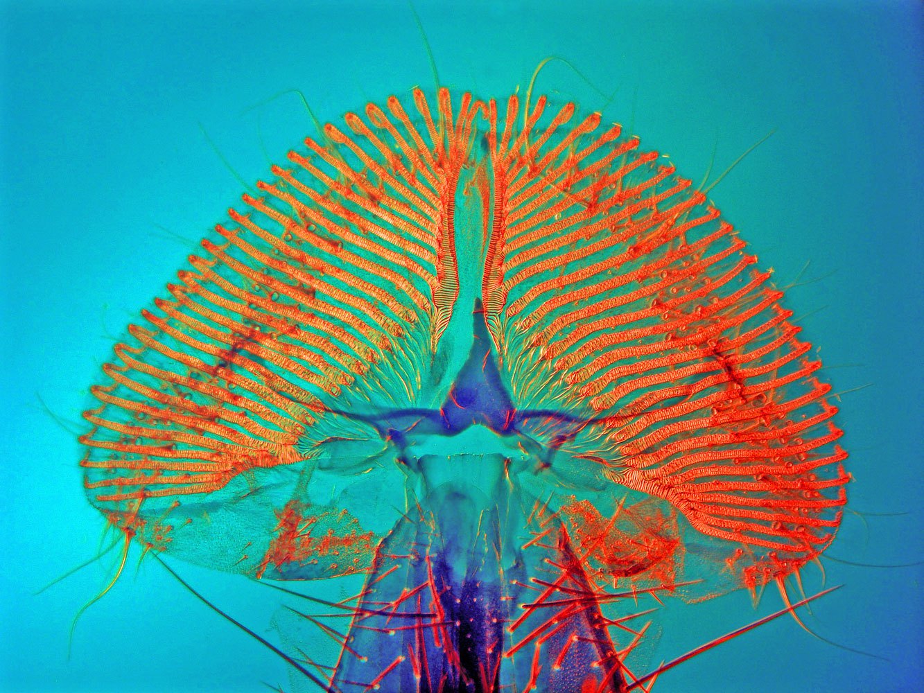
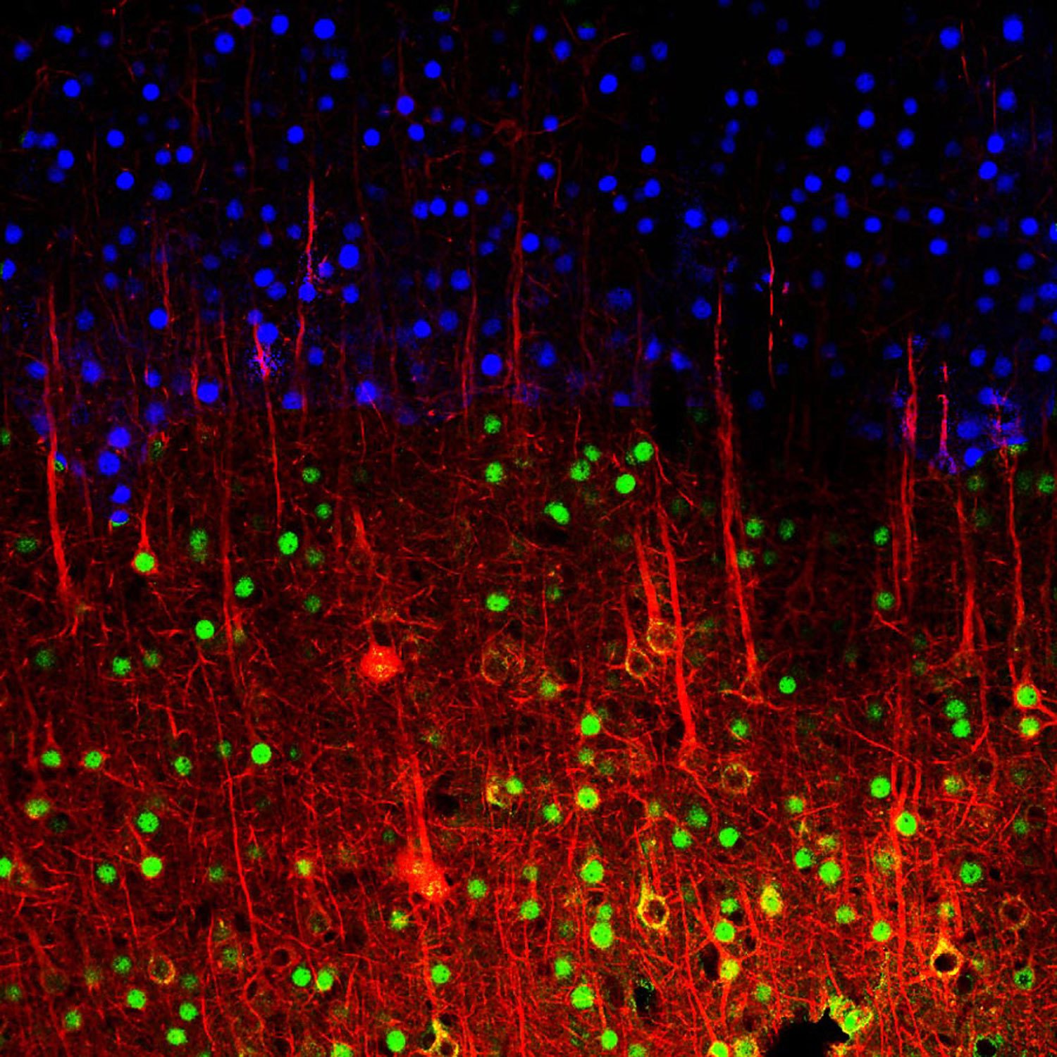
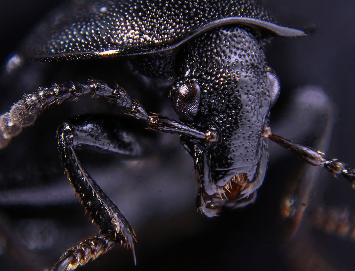
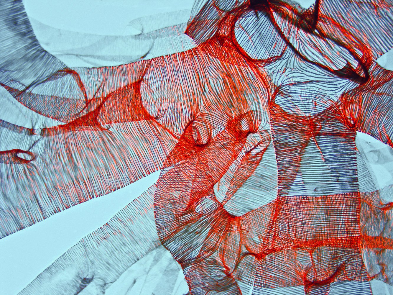
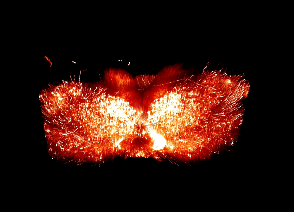
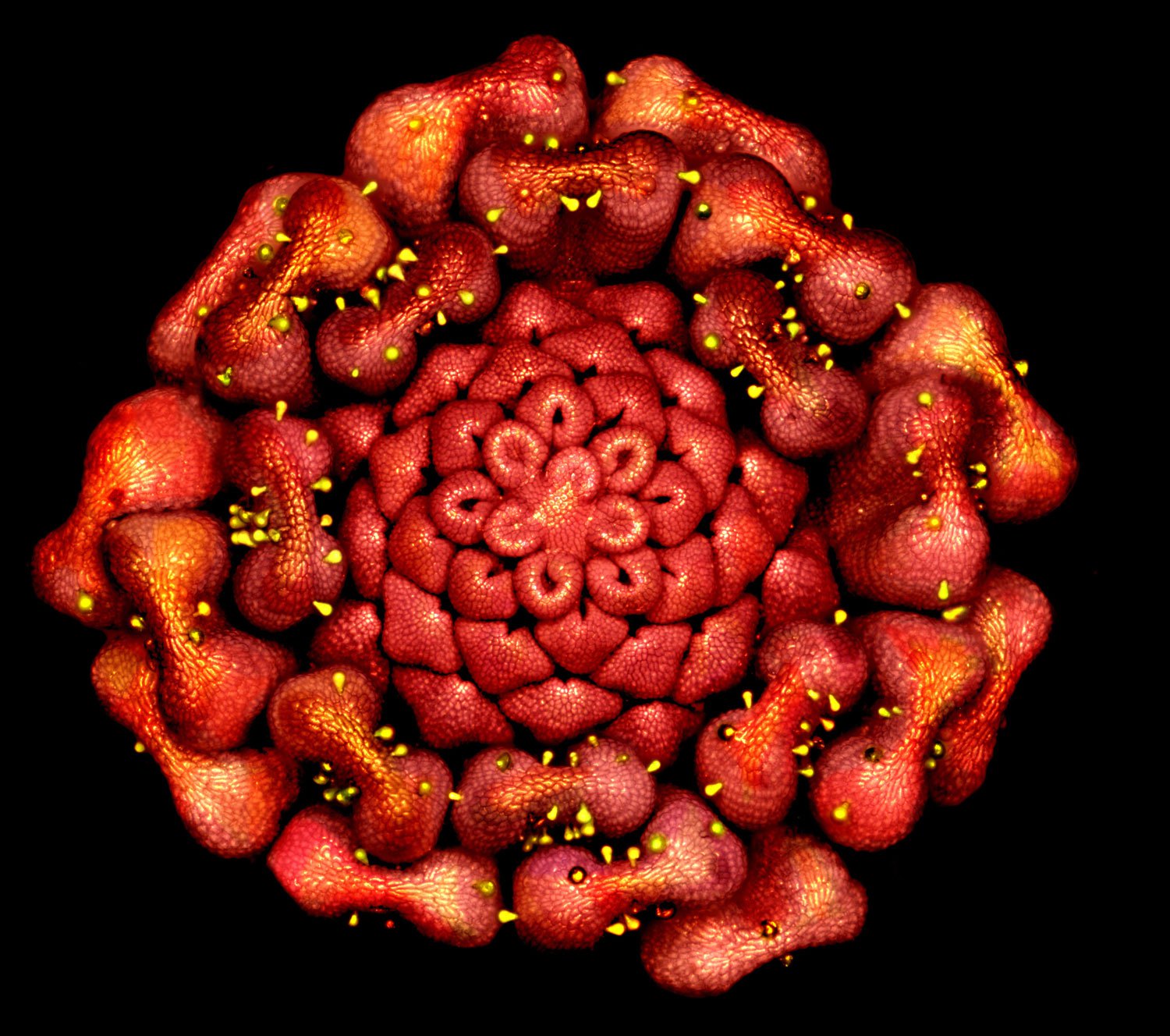
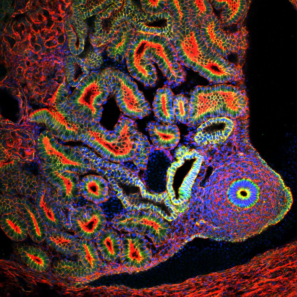
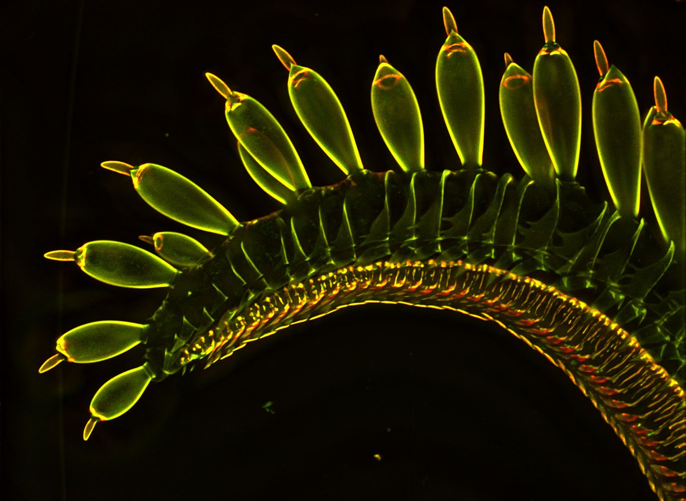
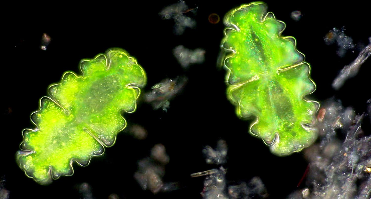
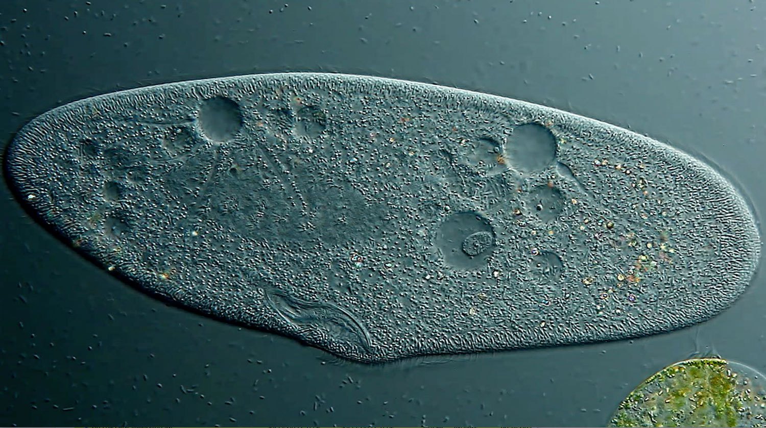
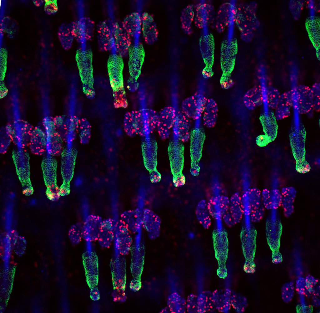
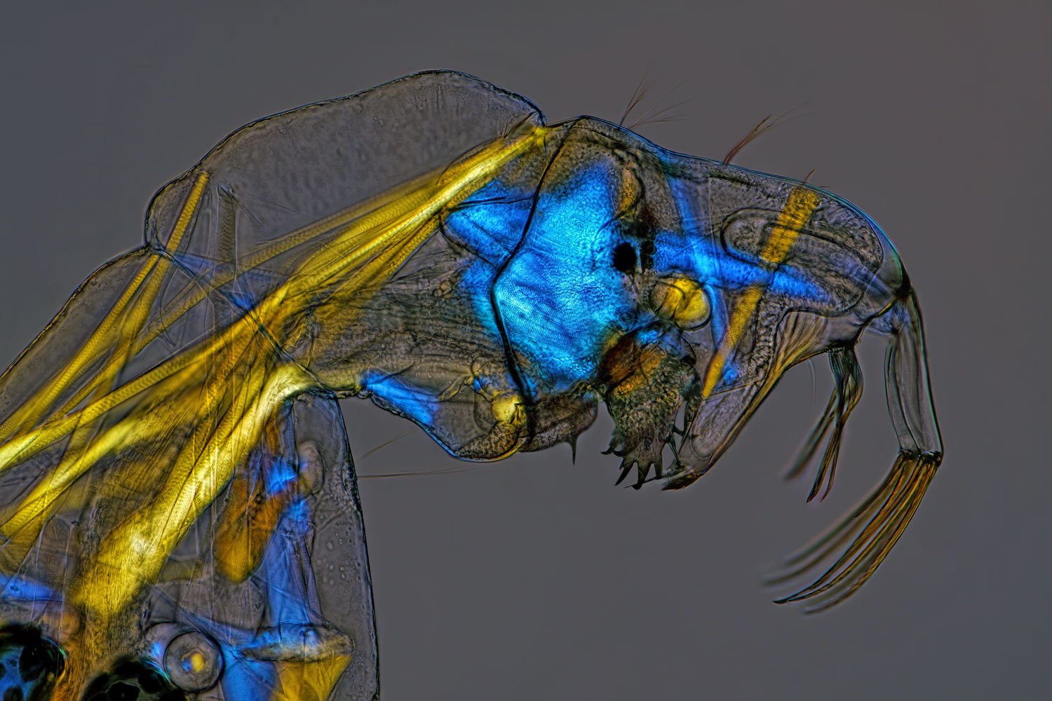
Tiny Beauties: Life's Smallest Wonders As Seen Through a Microscope
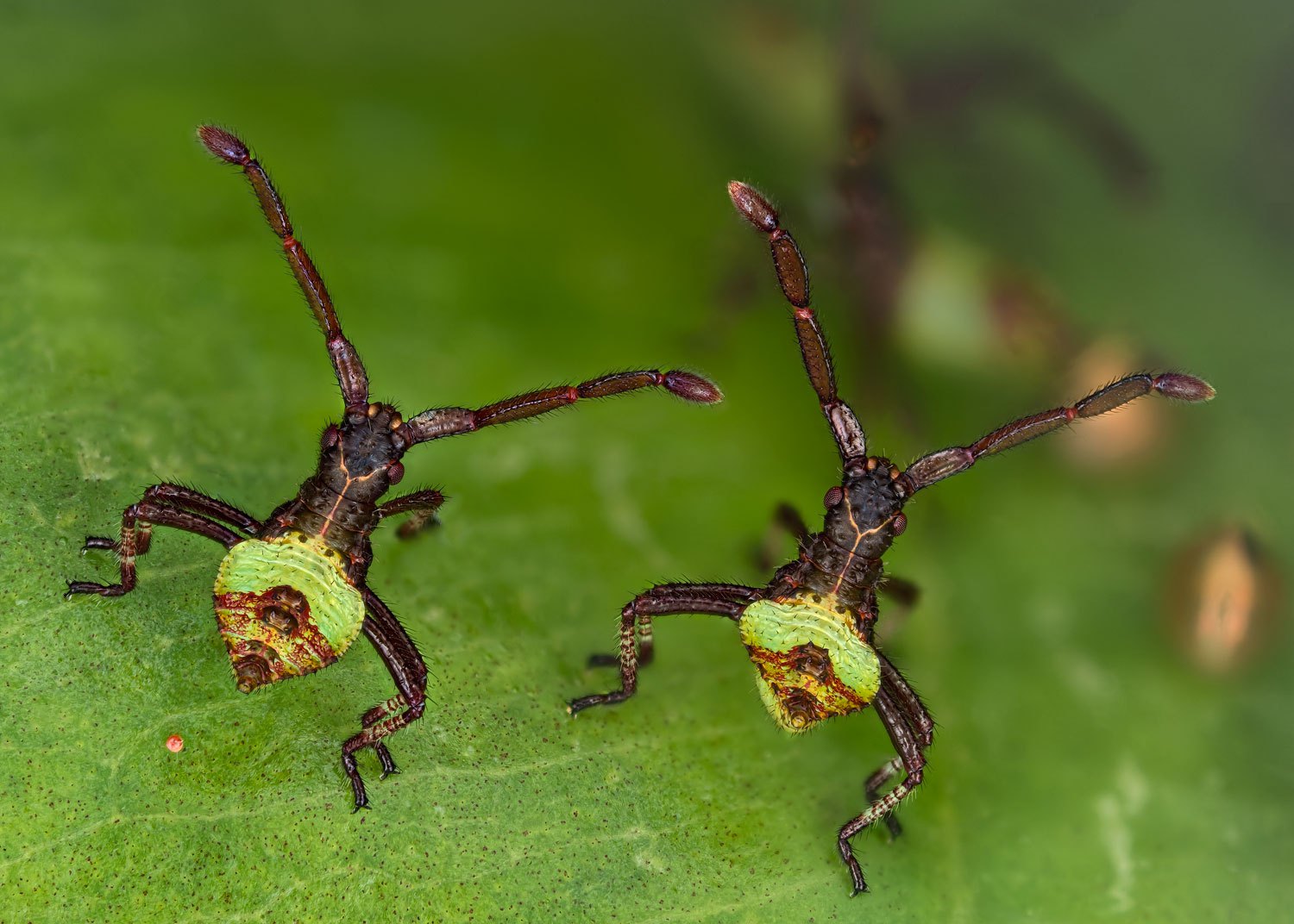
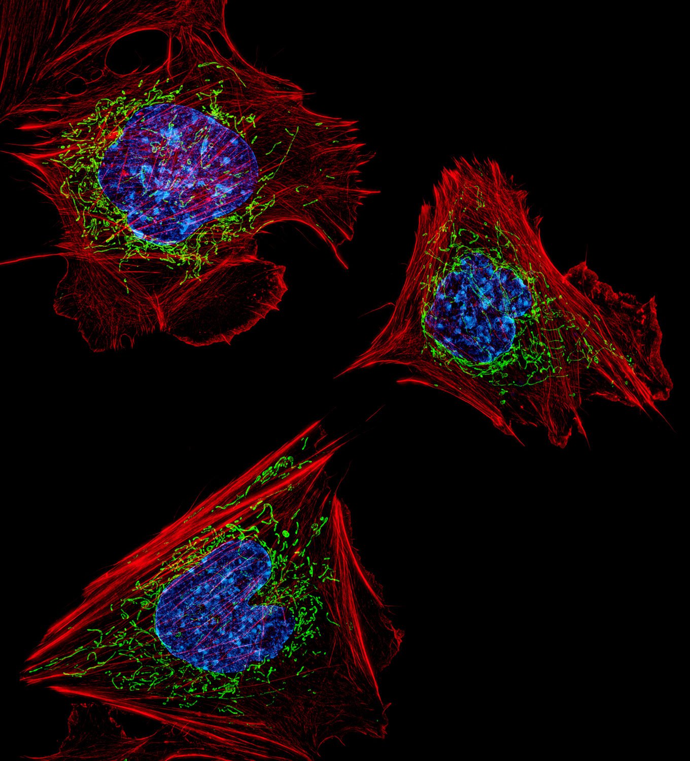
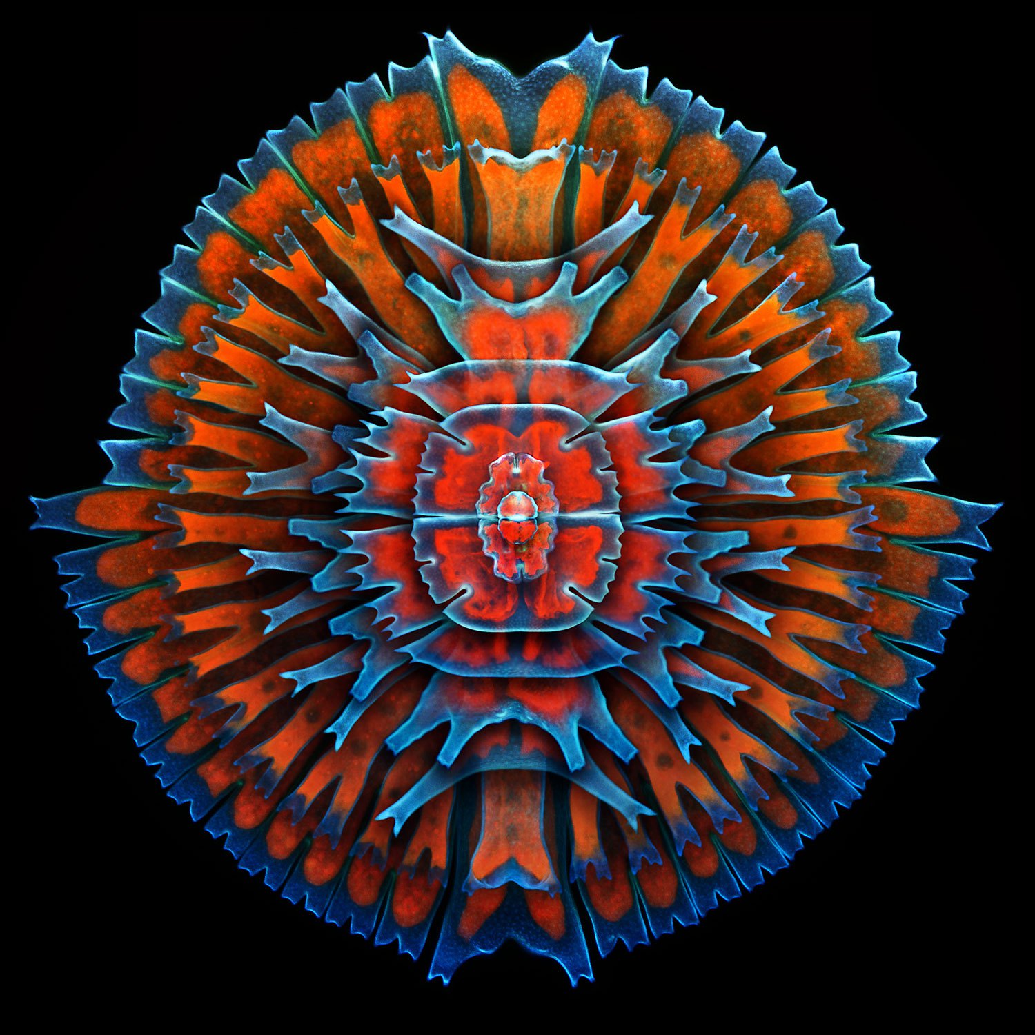
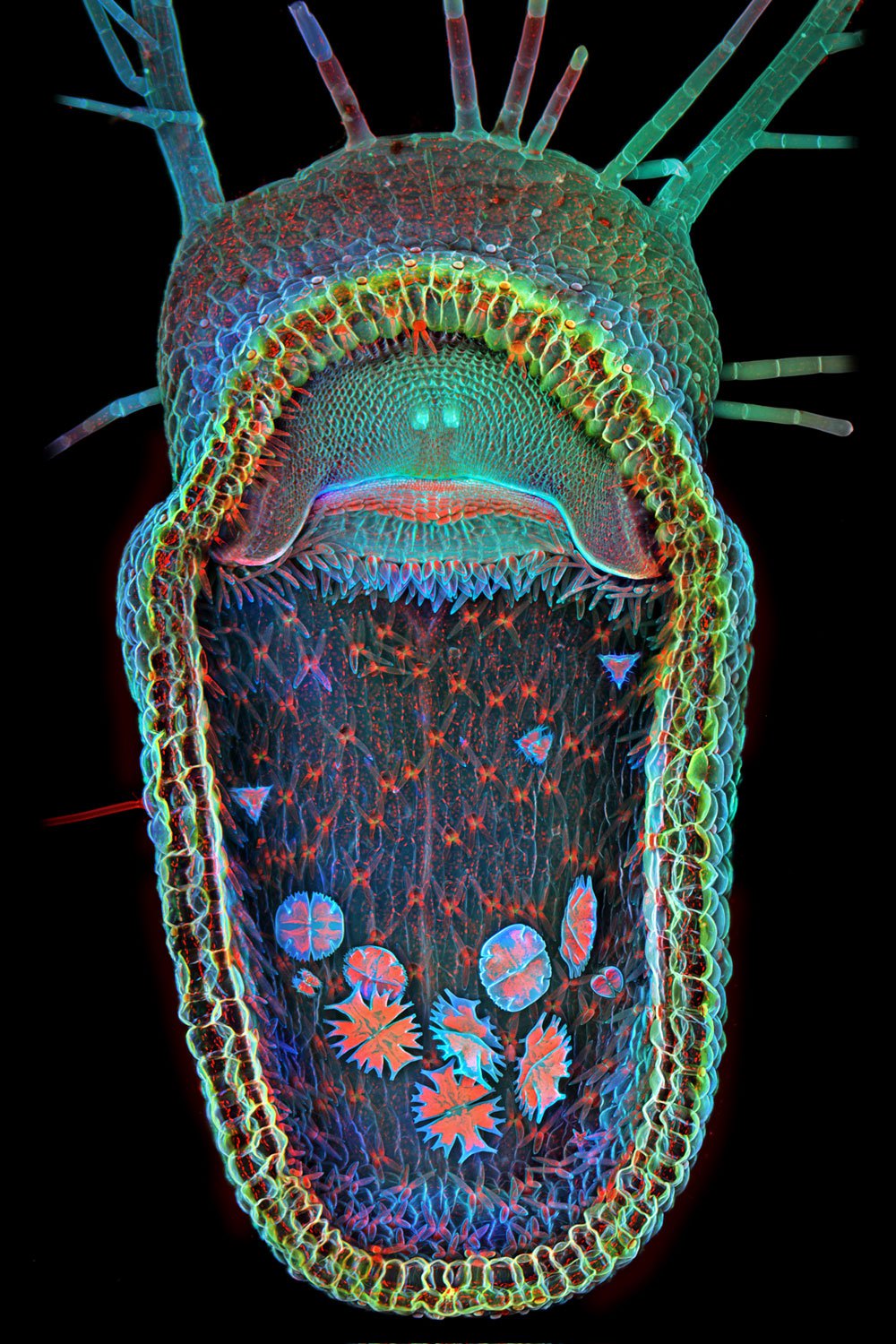
Honorable Mention: Proboscis of a blowfly.Michael Gibson—Olympus BioscapesHonorable Mention: Adult mouse cerebral cortex.Dr. Claudia Barros—Olympus BioscapesHonorable Mention: Black beetle.Pekka Honkakoski—Olympus BioscapesHonorable mention: Tracheae of a silkworm.Michael Gibson—Olympus BioscapesHonorable mention (Video Still): Axons in a mouse brainstem. Dr. Ali Ertürk—Olympus BioscapesHonorable Mention: Anemone flower.Masoumeh "Sahar" Khodaverdi—Olympus BioscapesHonorable Mention: Chick embryonic kidney.Dr. Poulomi Ray—Olympus BioscapesHonorable Mention: Tip of the proboscis of a Viceroy butterfly.Dr. Matthew S. Lehnert and Catherine P. Mulvane—Olympus BioscapesHonorable mention (Video Still): Cell division, movements and cytoplasmic streaming of the desmid Euastrum oblongum.Dr. Jens Hallfeldt—Olympus BioscapesHonorable Mention: Placental vasculature of a transgenic mouse embryo.Amanda Phillips-Yzaguirre—Olympus Bioscapes10th Place (Video Still): Paramecium, showing contactile vacuole and ciliary motion.Ralph Grimm—Olympus Bioscapes9th Place: Head and legs of a caddisfly larva.Fabrice Parais—Olympus Bioscapes8th Place: Mouse tail stained for the K15 (green) hair follicle stem cell marker as well as Ki67 (red), which marks proliferating cells.Dr. Yaron Fuchs—Olympus Bioscapes7th Place: Phantom midge larva (Chaoborus).Charles Krebs—Olympus Bioscapes6th Place: Gonocerus acuteangulatus, two hours old.Kurt Wirz—Olympus Bioscapes5th Place: Mouse embryonic fibroblasts showing the actin filaments (red) and DNA (blue).Dr. Dylan Burnette—Olympus Bioscapes4th Place: Stained transverse section of a lily flower bud.Spike Walker—Olympus Bioscapes3rd Place: A composite image showing a collection of single-cell fresh water algae, desmids.Dr. Igor Siwanowicz—Olympus Bioscapes2nd Place: A lateral view of a black mastiff bat embryo (Molossus rufus).Dorit Hockman—Olympus Bioscapes1st Place: Open trap of aquatic carnivorous plant, humped bladderwort (Utricularia gibba).Dr. Igor Siwanowicz—Olympus Bioscapes