Even though KSNT reporter Katya Leick was recording her stand-up from a tank at Fort Riley near Topeka, Kansas, surrounded by Army personnel in fatigues, it seemed like nothing could protect her from an ambush of pestering cicadas.
In video that, fortunately for Leick, was not live, she can be seen swatting the insects away and shrieking when they stuck to her face. Also fortunately for Leick — and anyone else who ends up in this situation — experts say the insects are harmless.
Tiny Beauties: Life's Smallest Wonders As Seen Through a Microscope
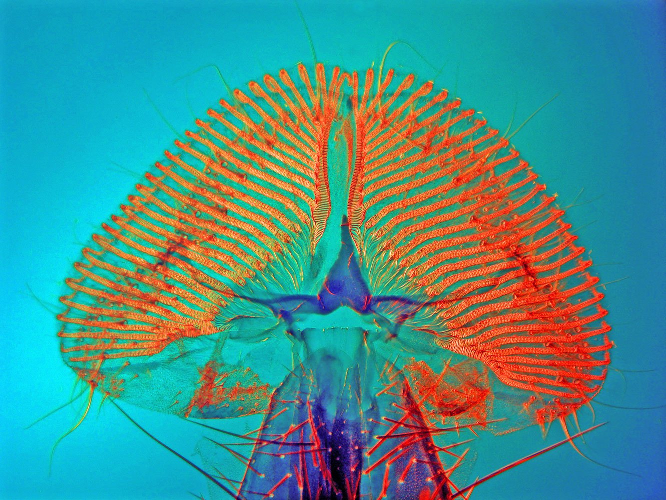
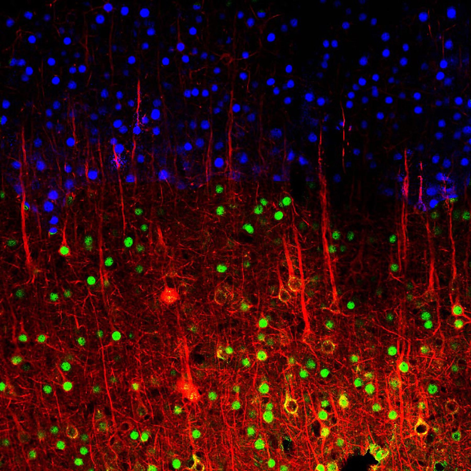
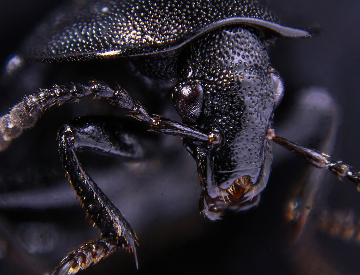
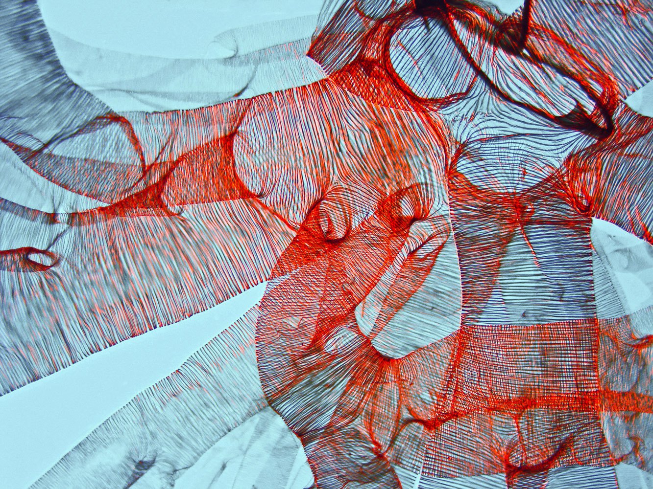
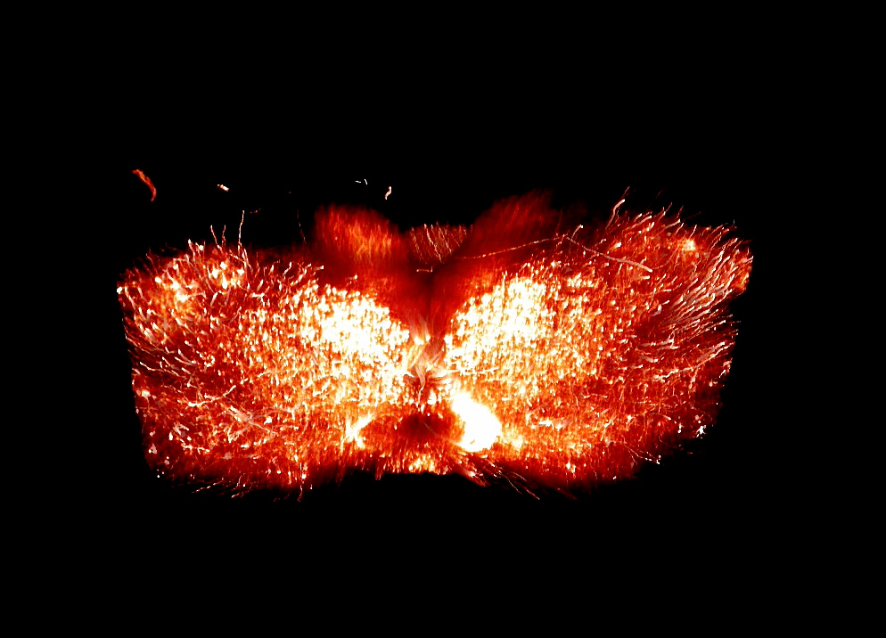
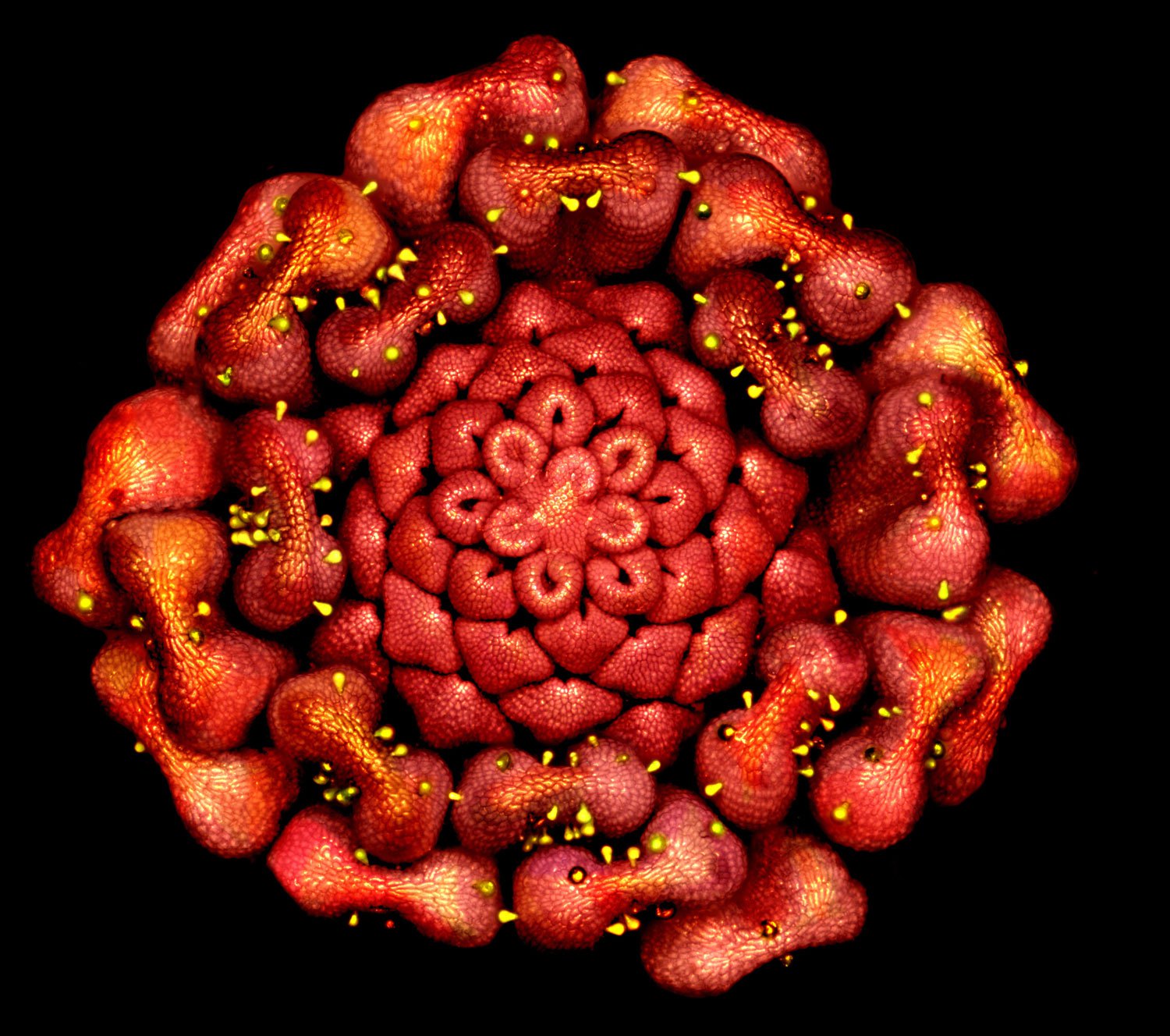
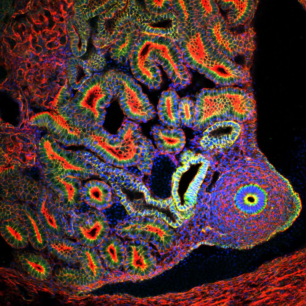
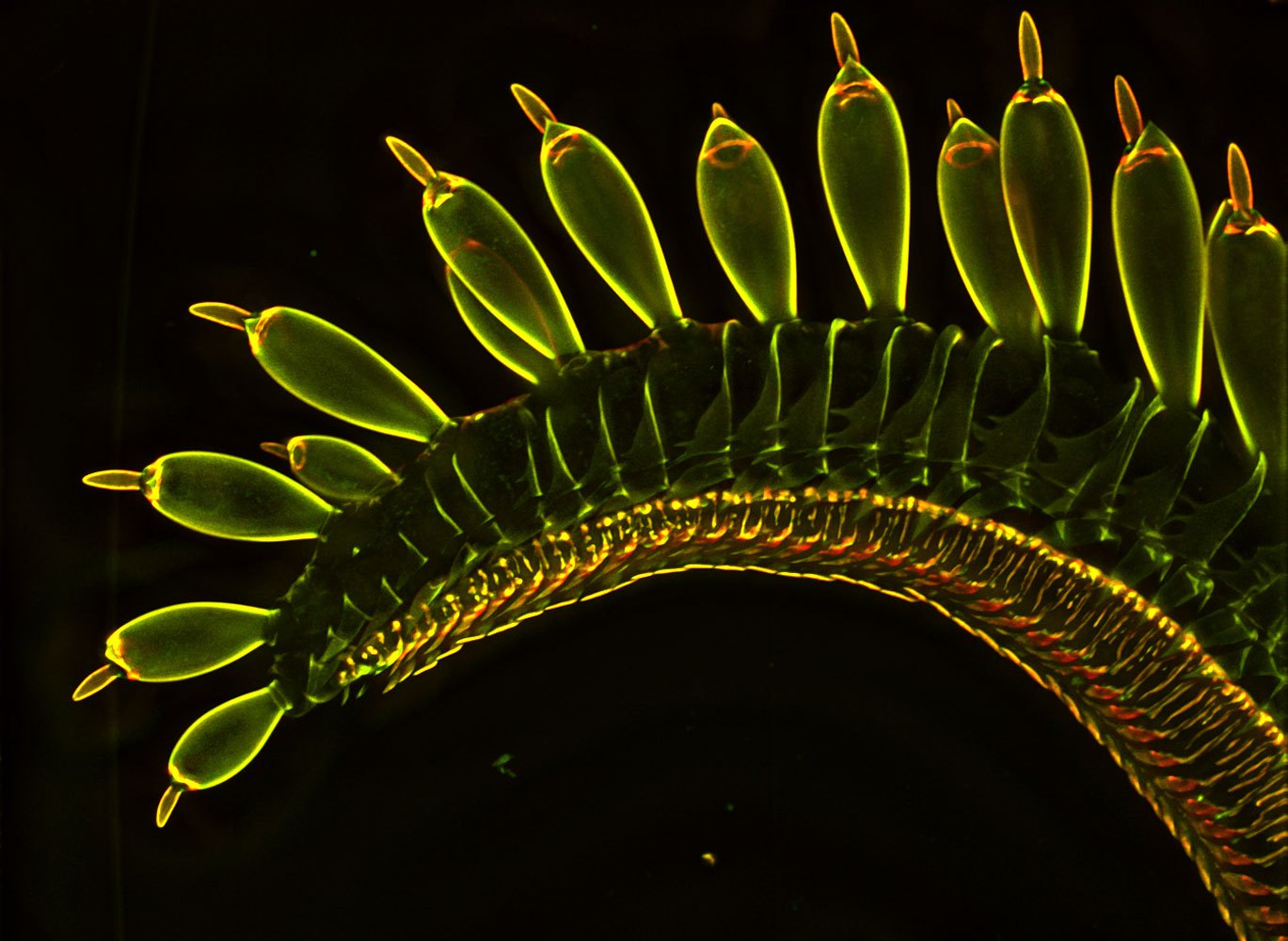
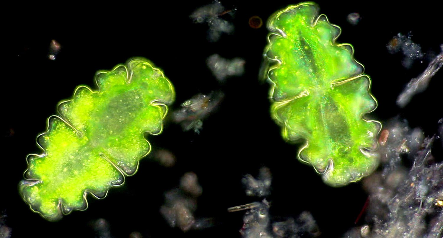
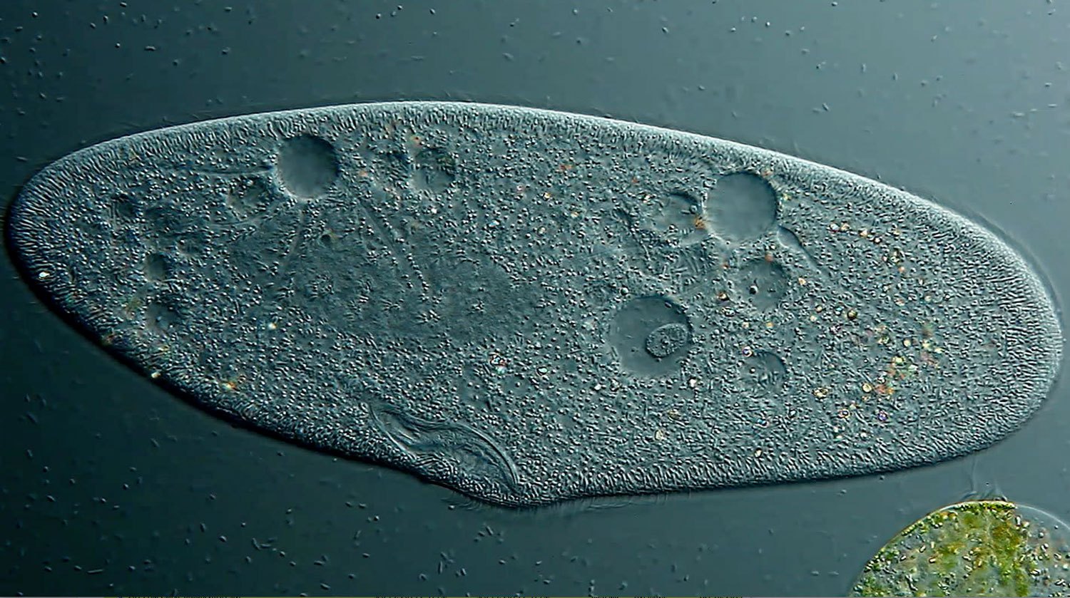
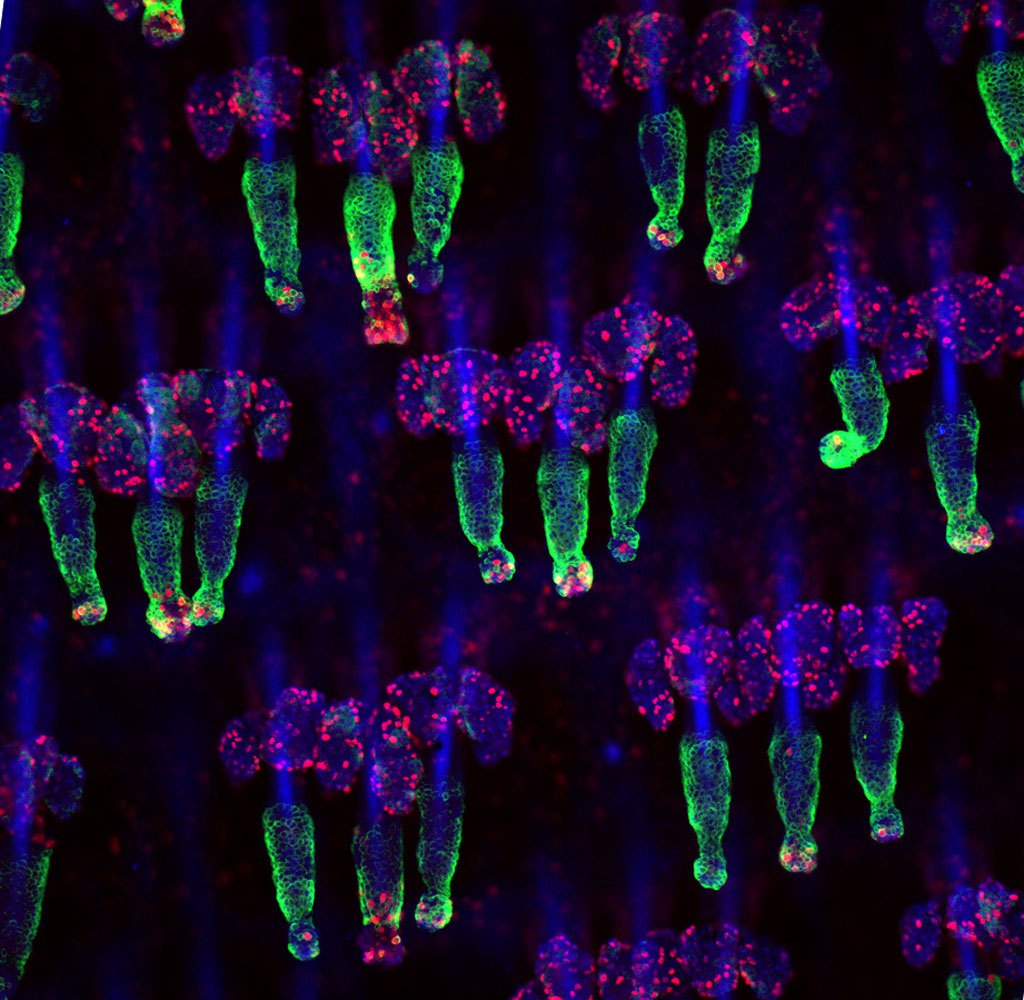
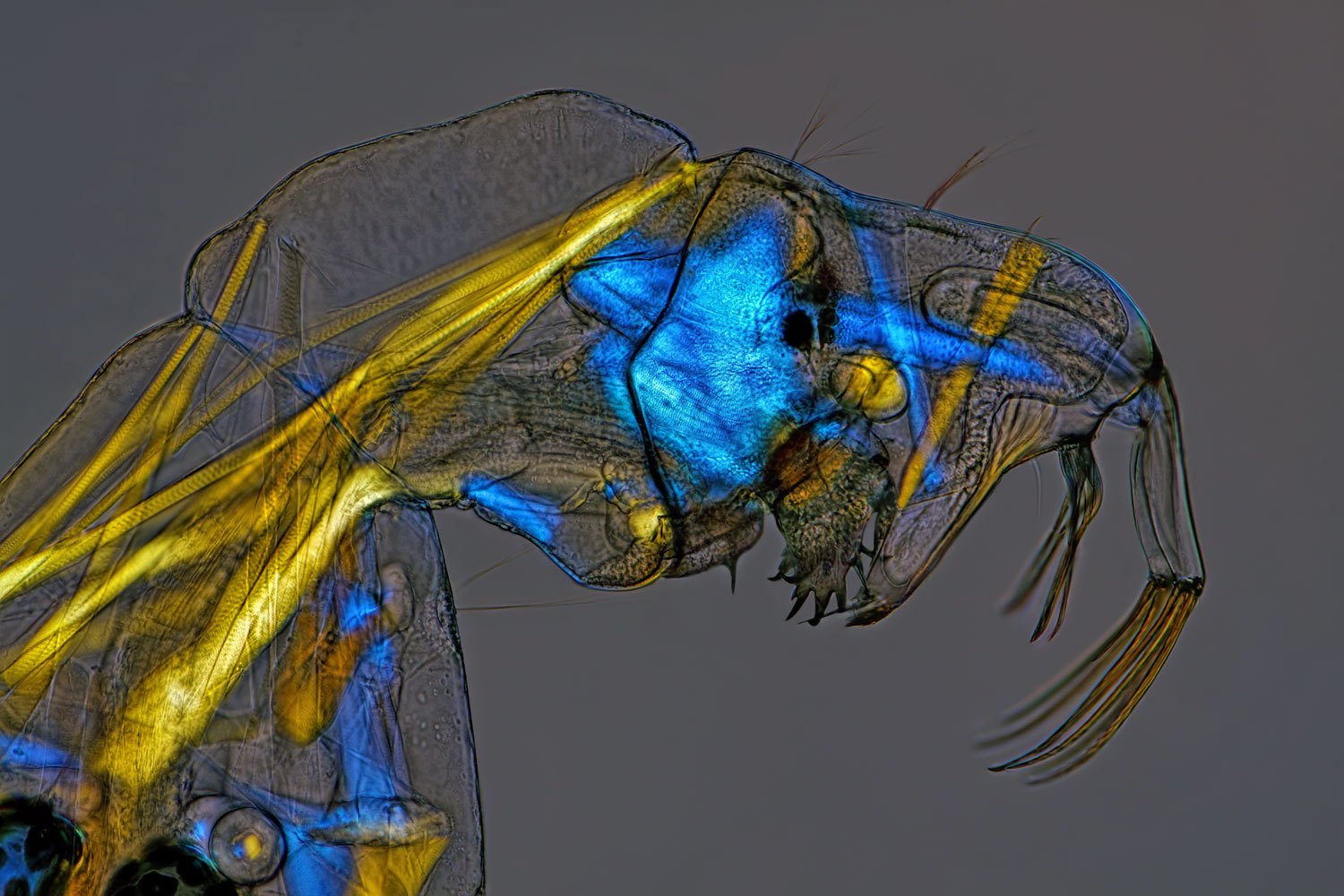
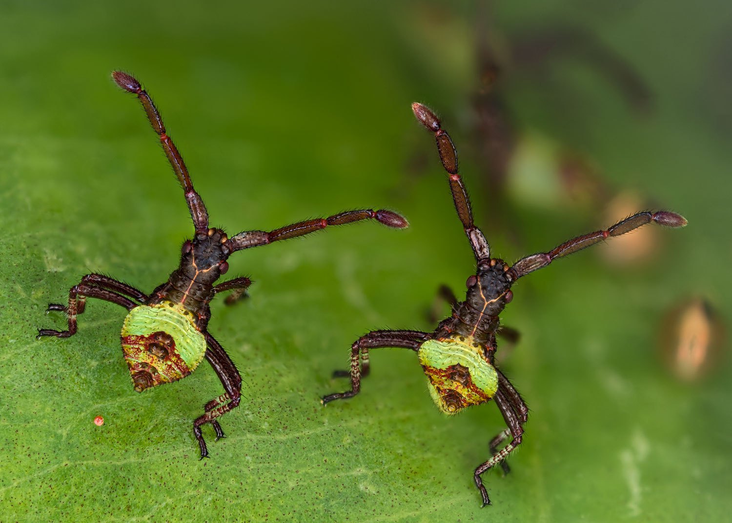
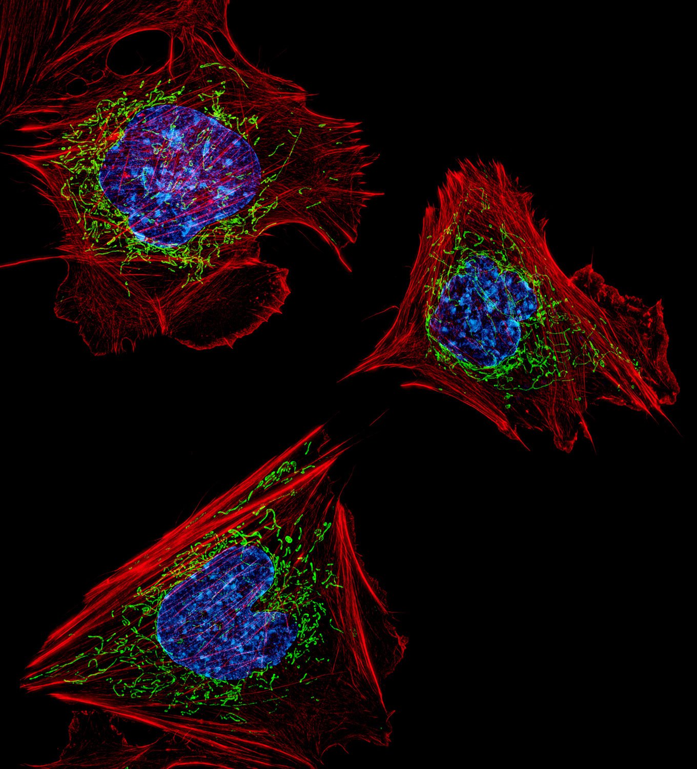
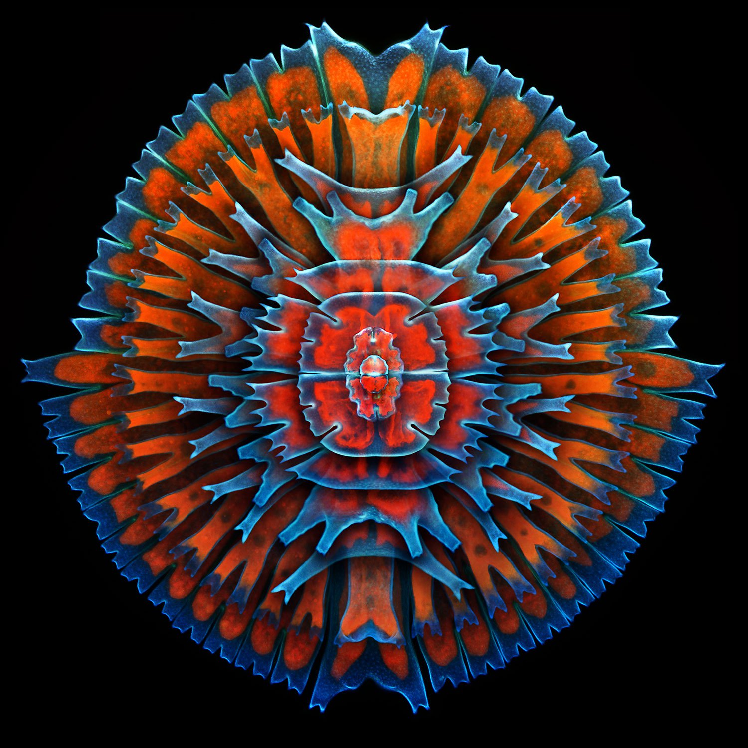
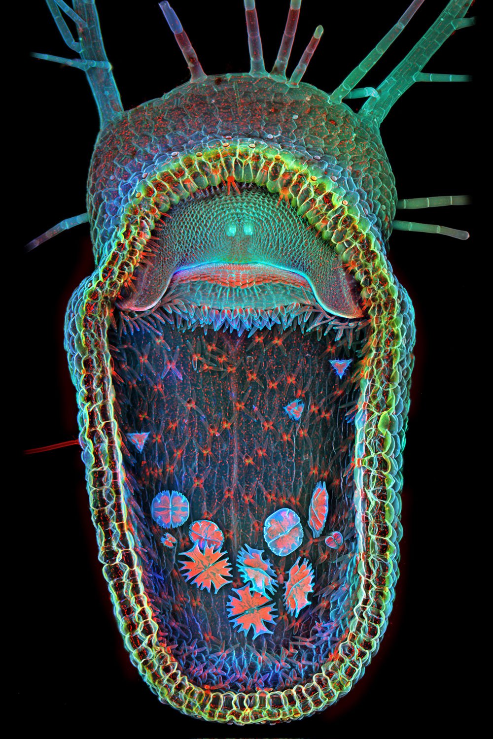
Honorable Mention: Proboscis of a blowfly.Michael Gibson—Olympus BioscapesHonorable Mention: Adult mouse cerebral cortex.Dr. Claudia Barros—Olympus BioscapesHonorable Mention: Black beetle.Pekka Honkakoski—Olympus BioscapesHonorable mention: Tracheae of a silkworm.Michael Gibson—Olympus BioscapesHonorable mention (Video Still): Axons in a mouse brainstem. Dr. Ali Ertürk—Olympus BioscapesHonorable Mention: Anemone flower.Masoumeh "Sahar" Khodaverdi—Olympus BioscapesHonorable Mention: Chick embryonic kidney.Dr. Poulomi Ray—Olympus BioscapesHonorable Mention: Tip of the proboscis of a Viceroy butterfly.Dr. Matthew S. Lehnert and Catherine P. Mulvane—Olympus BioscapesHonorable mention (Video Still): Cell division, movements and cytoplasmic streaming of the desmid Euastrum oblongum.Dr. Jens Hallfeldt—Olympus BioscapesHonorable Mention: Placental vasculature of a transgenic mouse embryo.Amanda Phillips-Yzaguirre—Olympus Bioscapes10th Place (Video Still): Paramecium, showing contactile vacuole and ciliary motion.Ralph Grimm—Olympus Bioscapes9th Place: Head and legs of a caddisfly larva.Fabrice Parais—Olympus Bioscapes8th Place: Mouse tail stained for the K15 (green) hair follicle stem cell marker as well as Ki67 (red), which marks proliferating cells.Dr. Yaron Fuchs—Olympus Bioscapes7th Place: Phantom midge larva (Chaoborus).Charles Krebs—Olympus Bioscapes6th Place: Gonocerus acuteangulatus, two hours old.Kurt Wirz—Olympus Bioscapes5th Place: Mouse embryonic fibroblasts showing the actin filaments (red) and DNA (blue).Dr. Dylan Burnette—Olympus Bioscapes4th Place: Stained transverse section of a lily flower bud.Spike Walker—Olympus Bioscapes3rd Place: A composite image showing a collection of single-cell fresh water algae, desmids.Dr. Igor Siwanowicz—Olympus Bioscapes2nd Place: A lateral view of a black mastiff bat embryo (Molossus rufus).Dorit Hockman—Olympus Bioscapes1st Place: Open trap of aquatic carnivorous plant, humped bladderwort (Utricularia gibba).Dr. Igor Siwanowicz—Olympus Bioscapes