Why Mosquitoes Will Always Track You Down

2 minute read

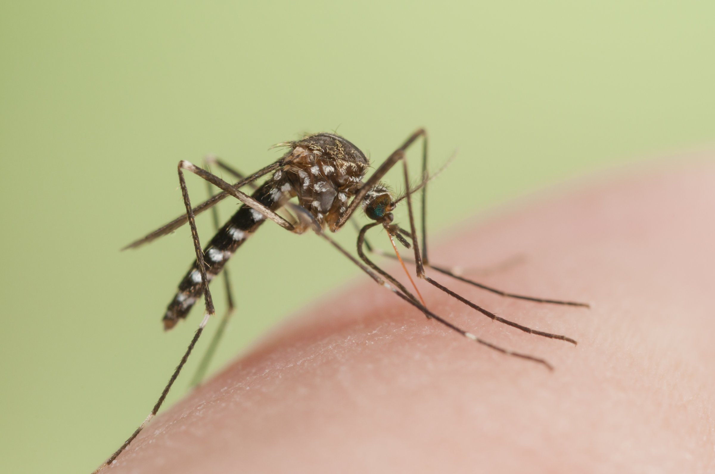
Ever wonder why mosquitoes seem drawn to your body like magnets? Scientists now understand the method the insects use to find their human prey, and it involves three steps.

According to new research published in the journal Current Biology, mosquitoes are attracted by the scent of CO2 — which is found in human breath. The insects can pick up on this trigger from a distance of 10 to 50 meters. Moving in closer, mosquitoes pick up on visual cues to find their target, an idea the researchers tested with a black spot on the floor of a wind tunnel. They can use sight to find humans from 5 to 15 meters. Finally, mosquitoes are attracted by the heat of the human body, which researchers confirmed with a heated glass panel that otherwise blended in with its surroundings. The insects can be drawn to heat from within a meter.

While scientists already knew that these three elements contributed to mosquitoes’ homing method, this is the first time they understand how all three parts work together.

[BBC]

See 40 Stunning Images Captured Through A Microscope
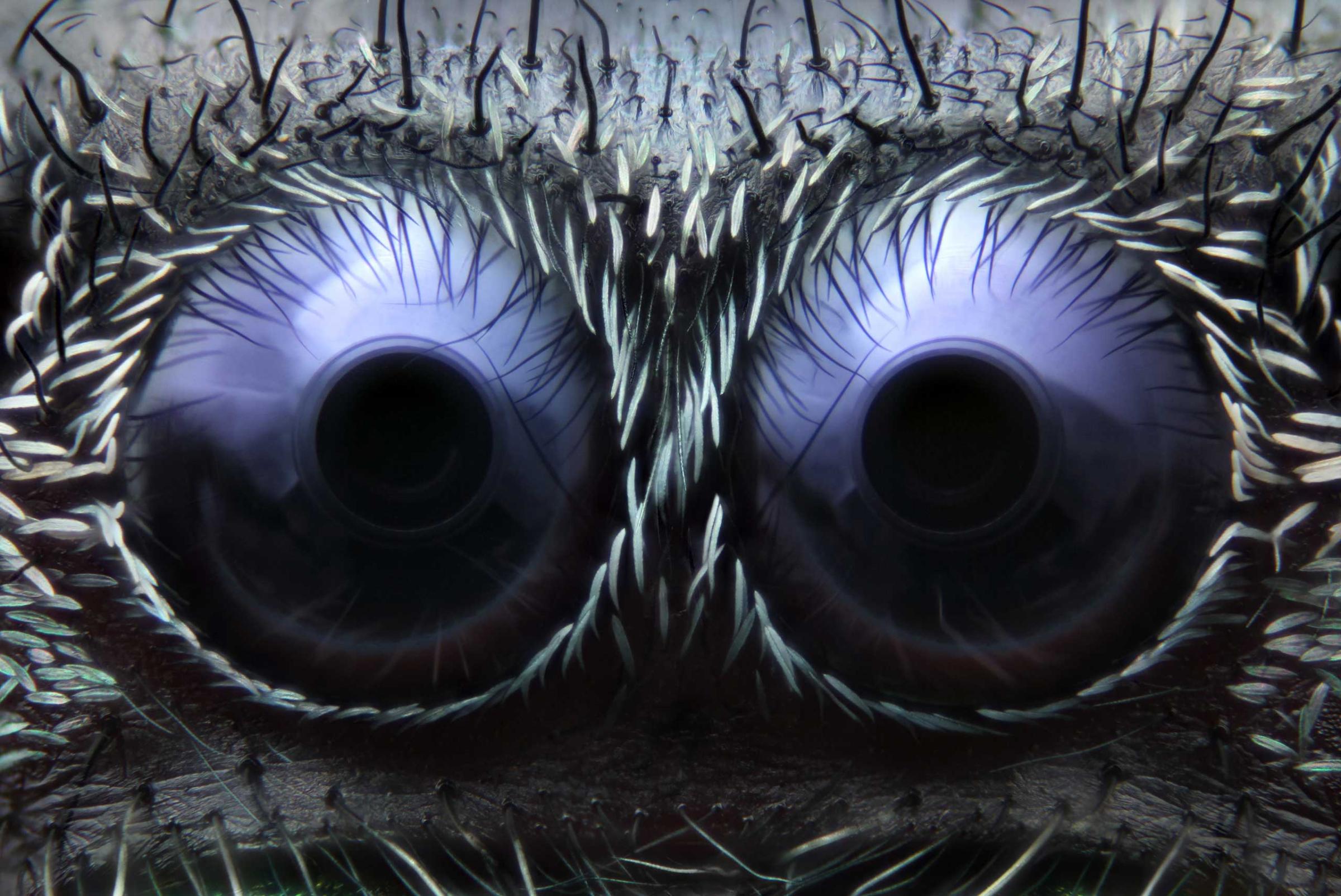
Jumping spider eyes at 20x magnification.Noah Fram-Schwartz
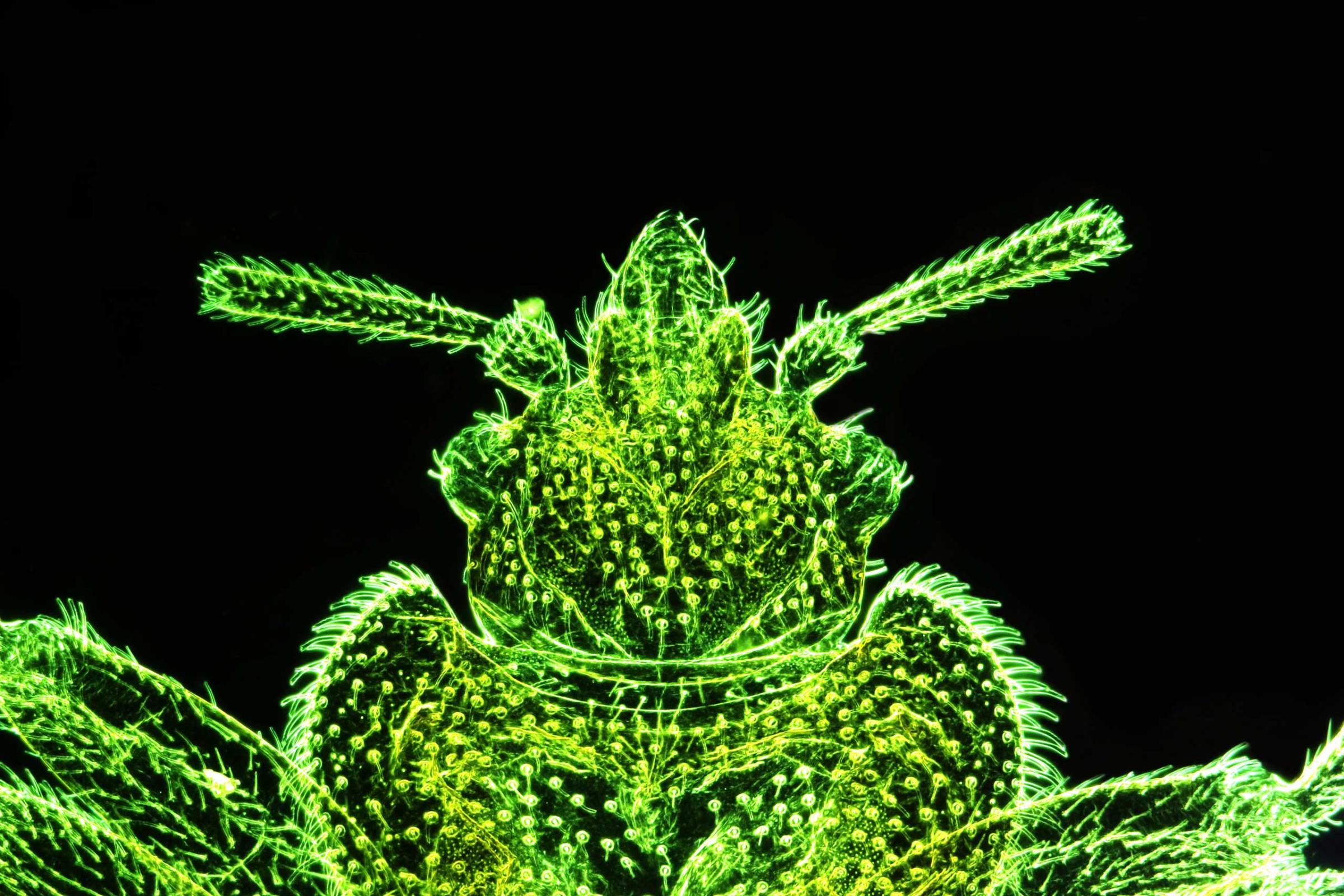
A bed bug at 50x magnification.Stefano Barone
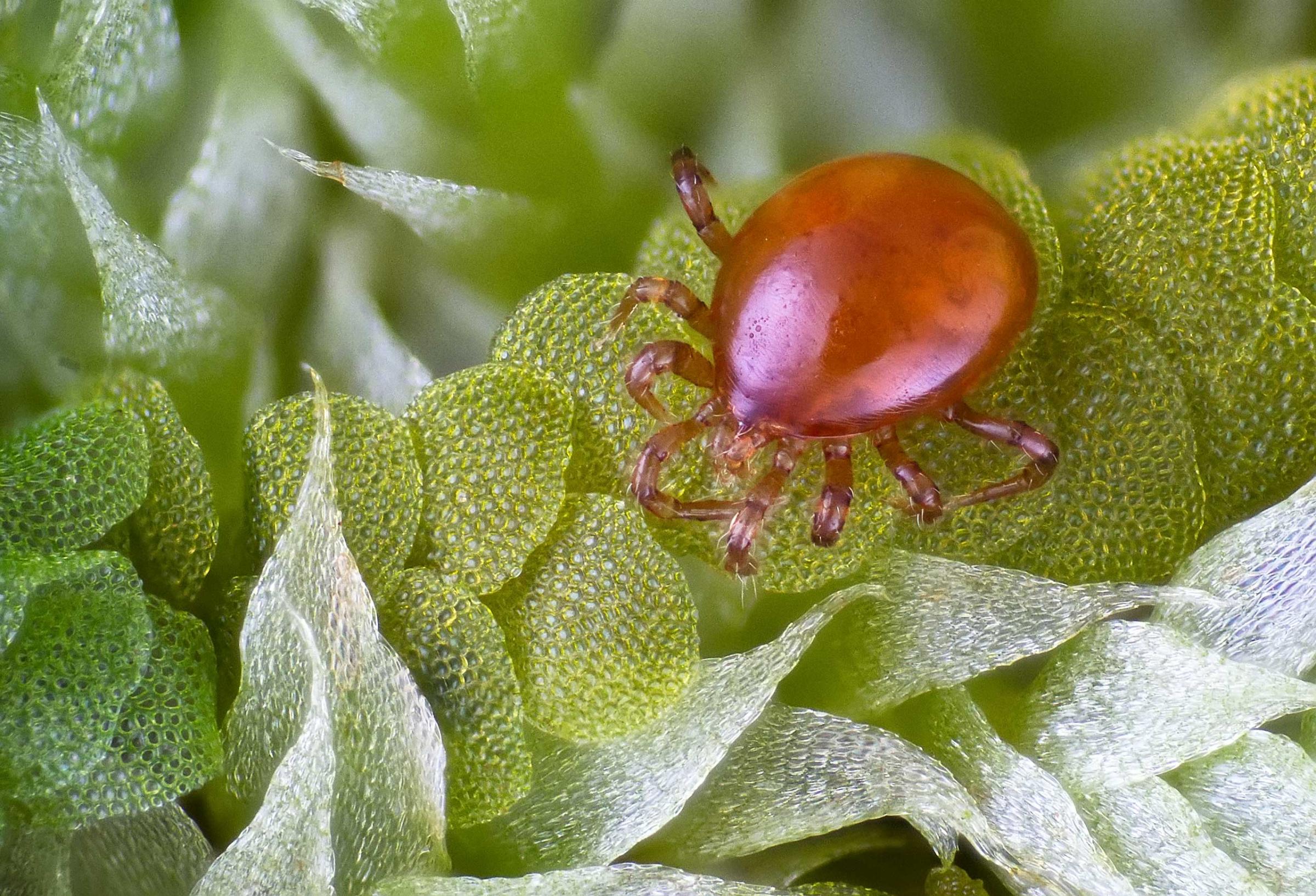
A mite in a forest at 10x magnification.José R. Almodóvar
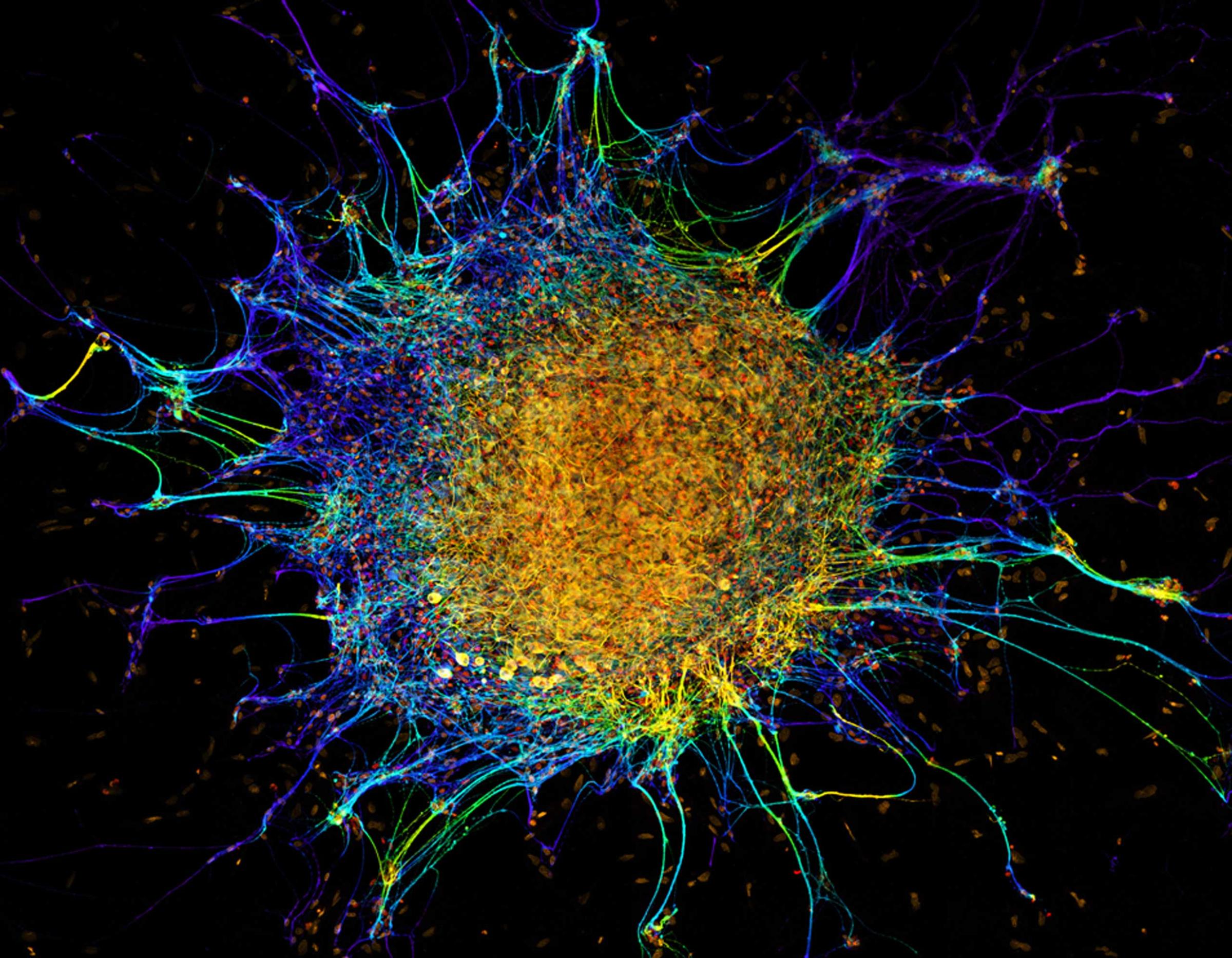
Cultured embryonic chicken dorsal root ganglia neuron explant at 60x magnification.Dr. Michael John Bridge
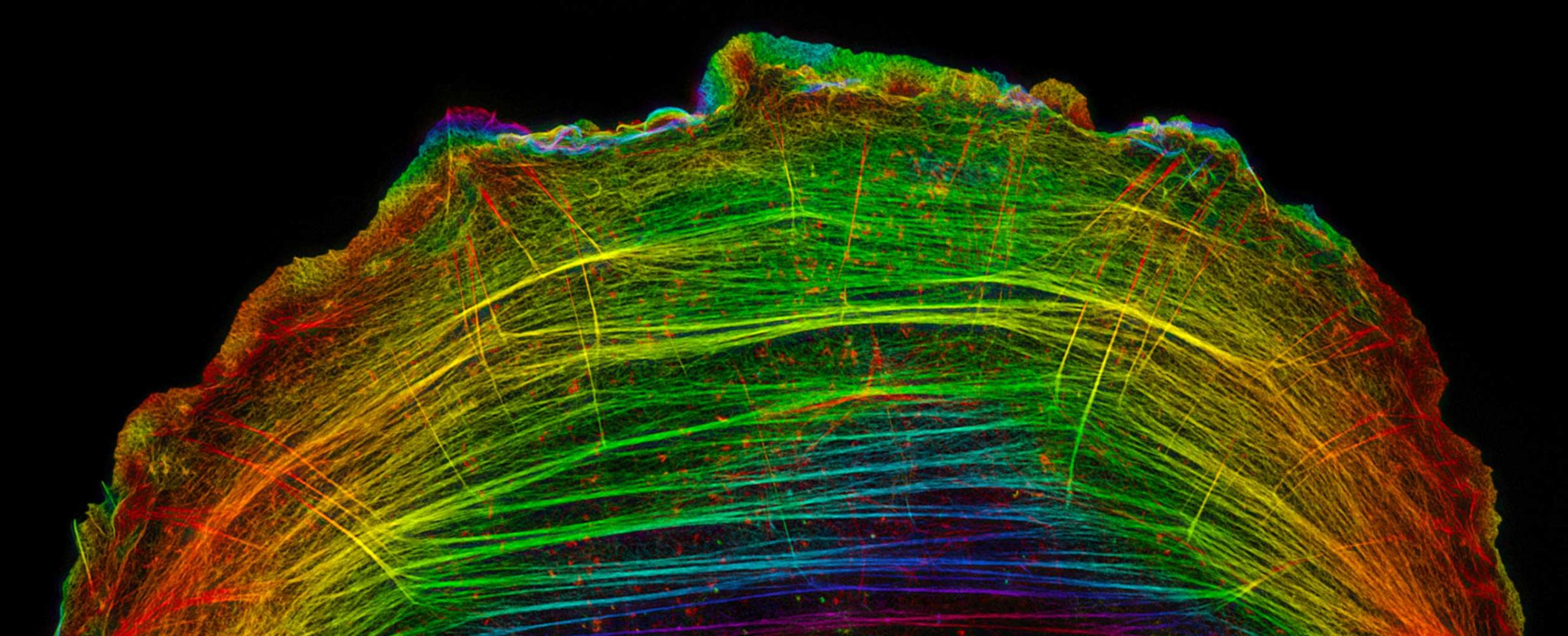
A crawling bone cancer cell
A crawling bone cancer cell at 8000x magnification.Dr. Dylan T. Burnette
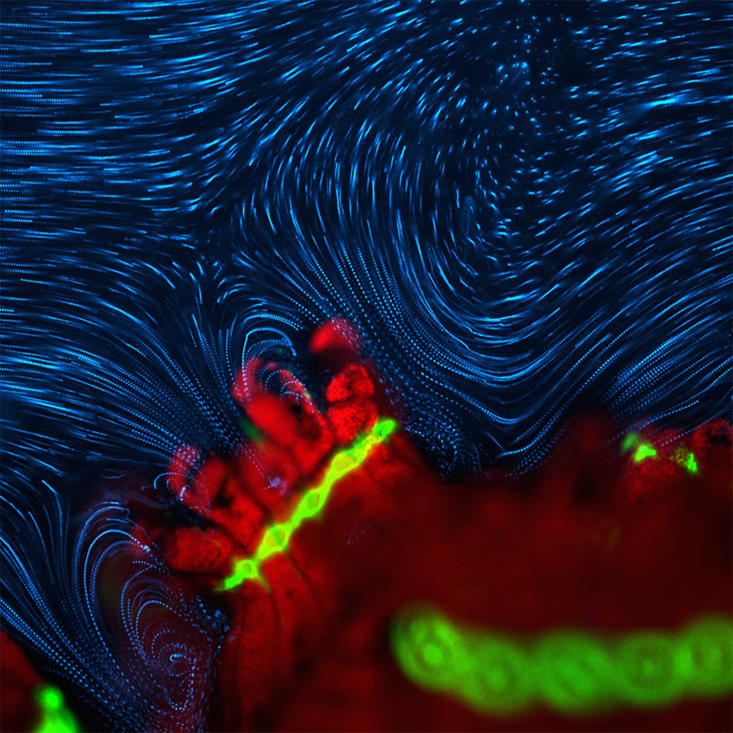
Active fluid flow around P. damicornis
Active fluid flow around P. damicornis at 4x magnificationDr. Douglas Brumley
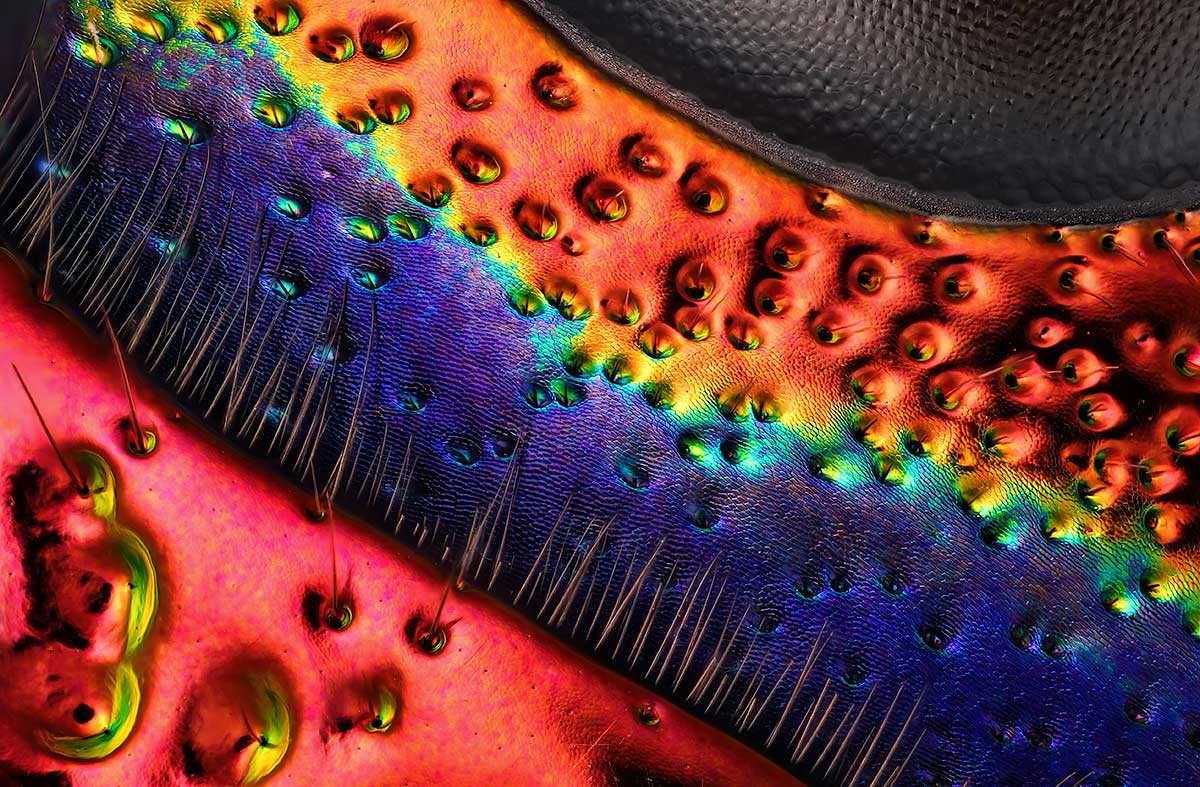
Chrysochroa buqueti (jewel beetle) carapace, near eye
A Chrysochroa buqueti (jewel beetle) at 45x magnification. Charles Krebs
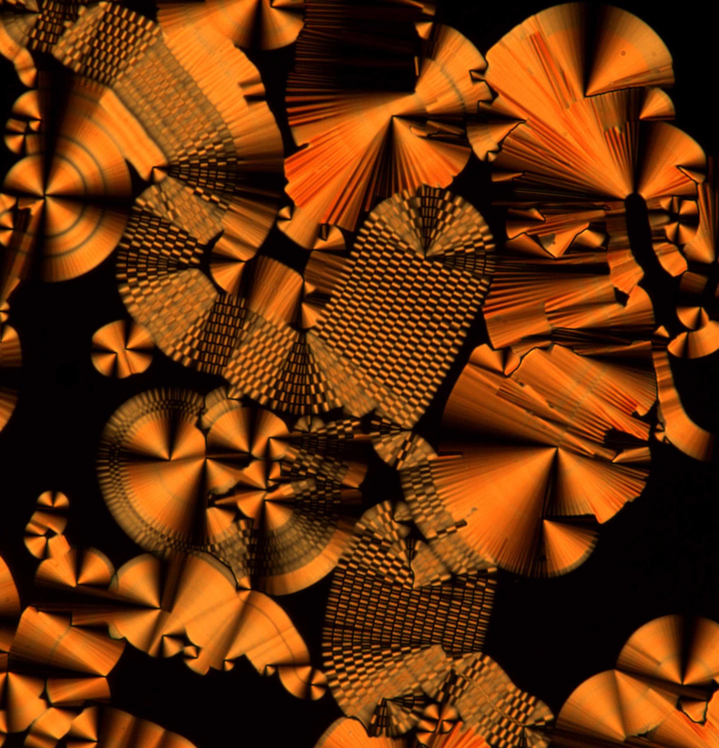
Focal conic-like domain with varying degrees of modulation and checkerboard patterns
Focal conic-like domain with varying degrees of modulation and checkerboard patterns at 40x magnification.Dr. Rajdeep Deb
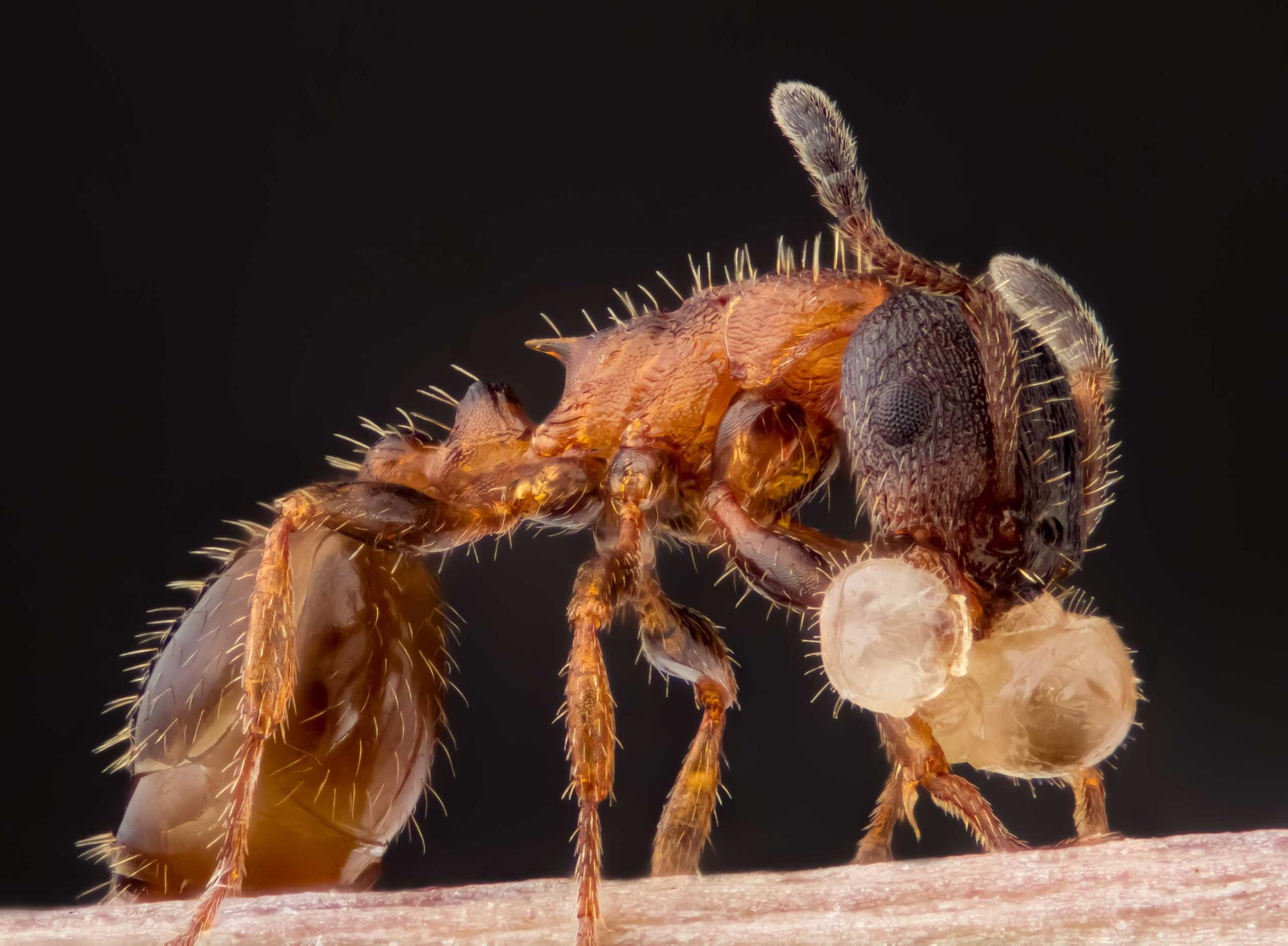
Leptothorax acervorum (ant) carrying its larva
ALeptothorax acervorum (ant) carrying its larva at 5x magnification.Geir Drange
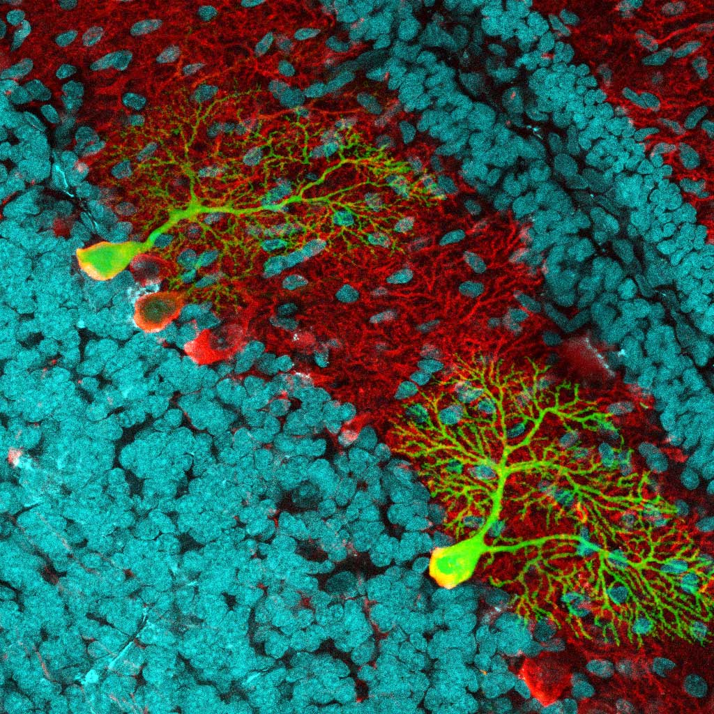
Sagittal brain slice showing cell nuclei (cyan) and Purkinije cells (red) expressing EGFP
A sagittal brain slice showing cell nuclei at 40x magnification.Dr. Marco Dal Maschio
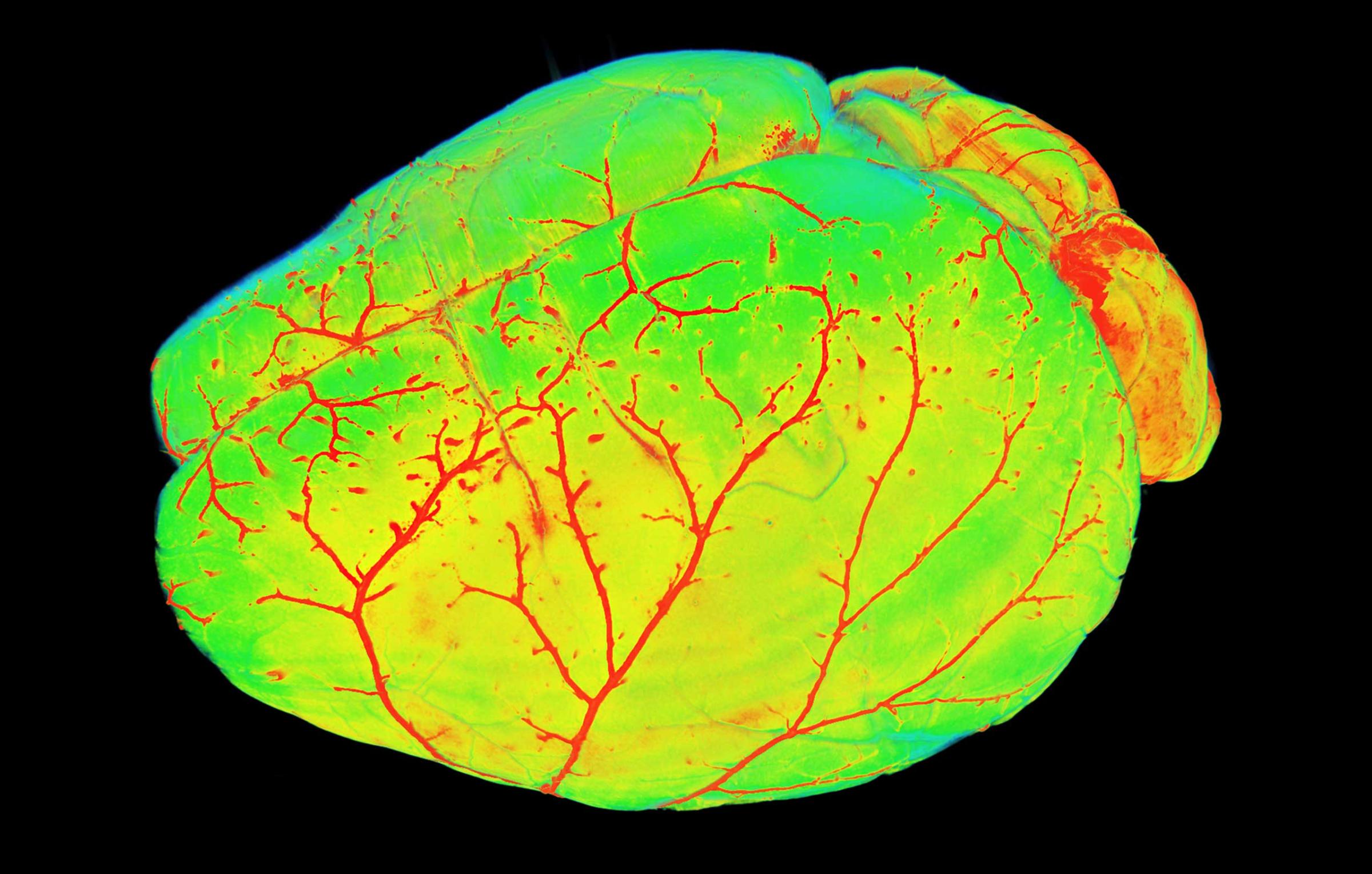
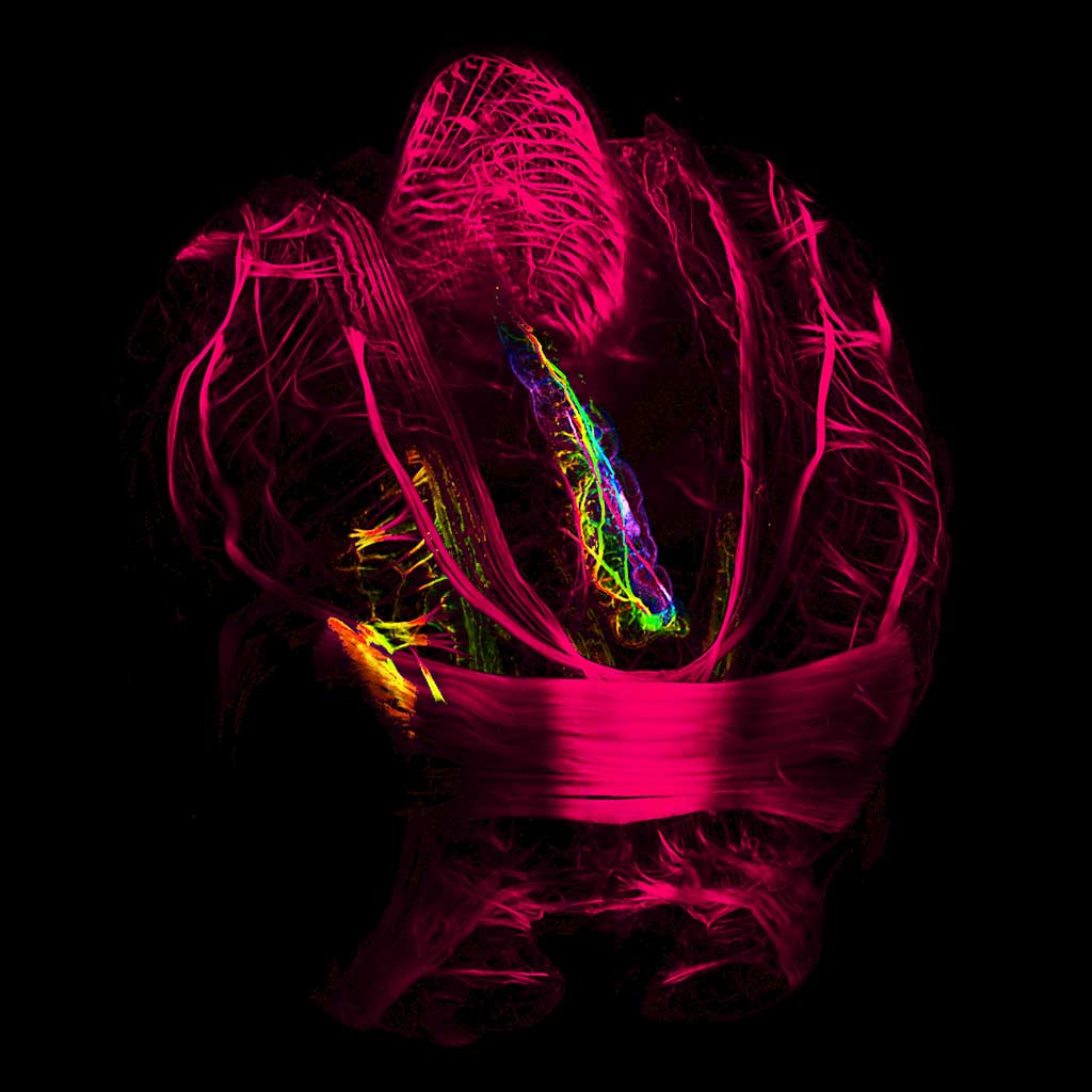
Mouse brain vasculature
A mouse brain vasculature at 2x magnification.Dr. Ali Erturk
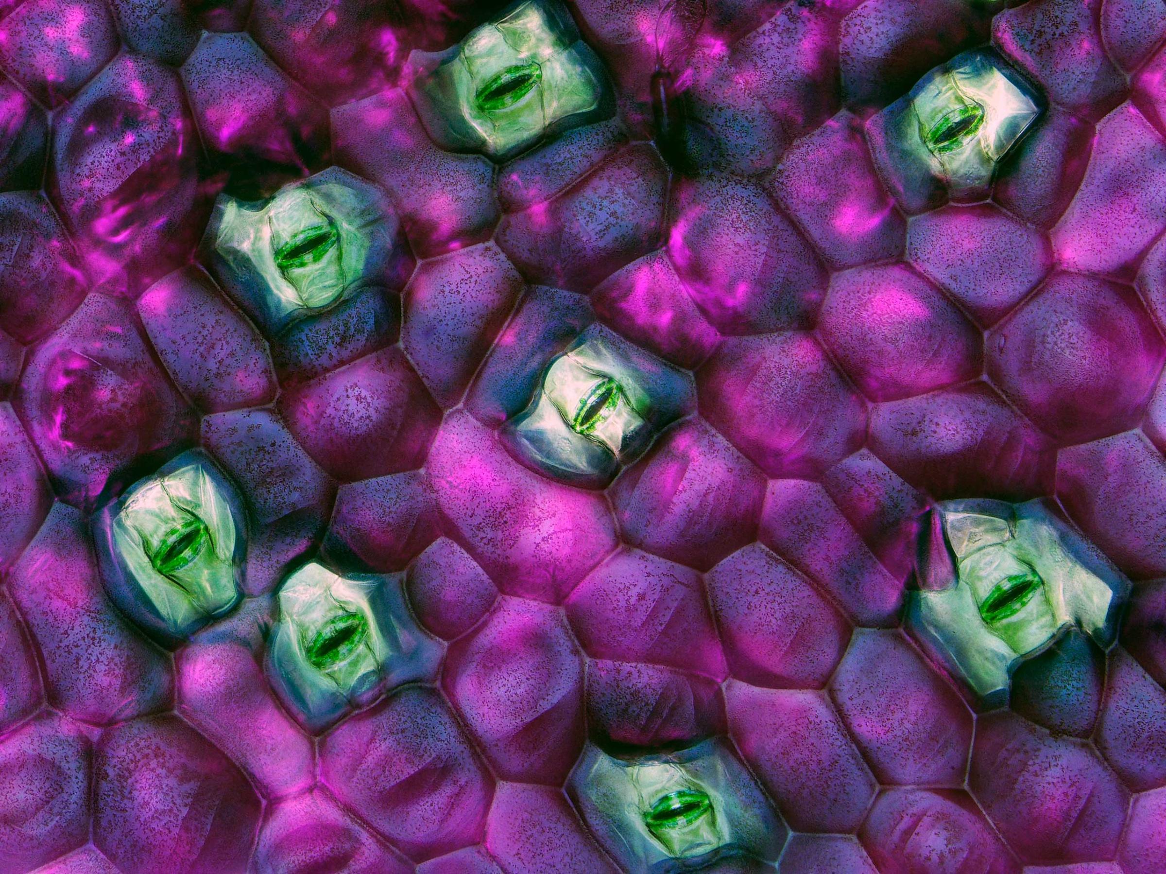
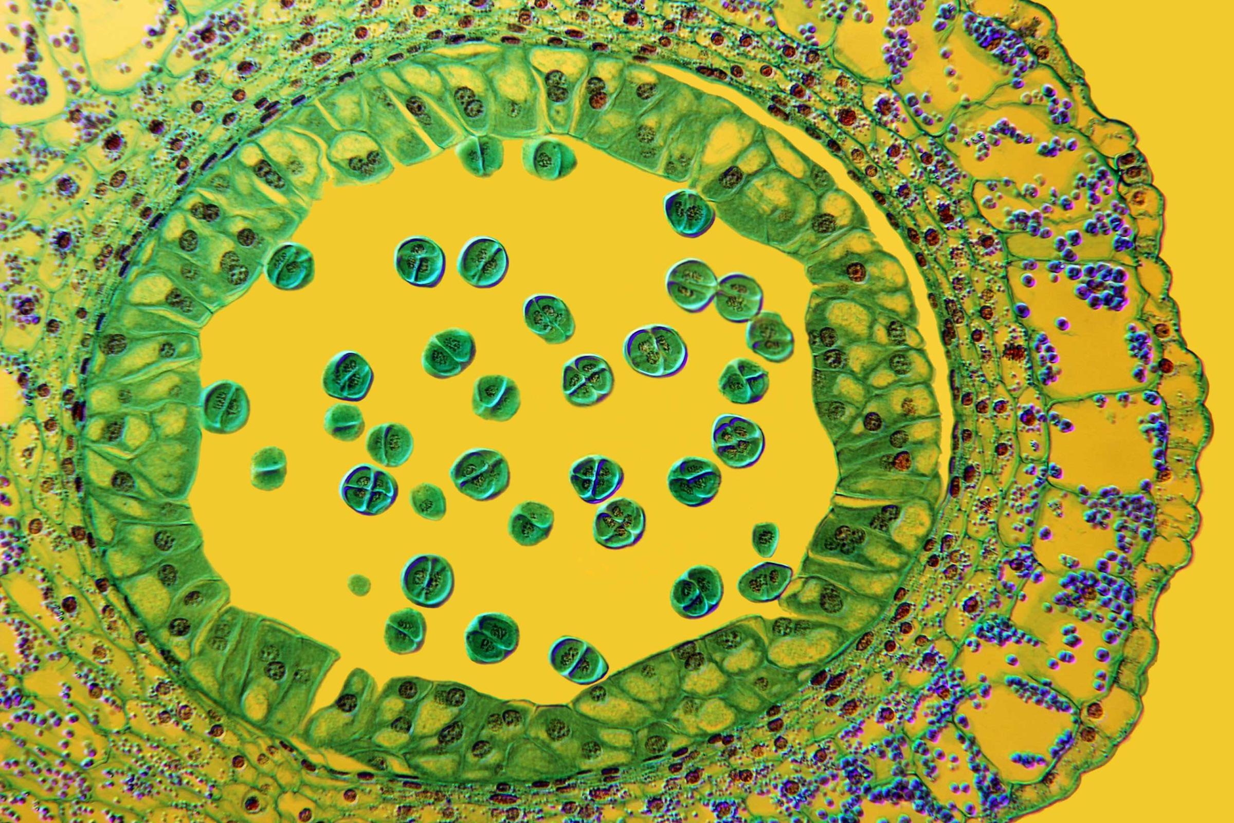
Tradescantia zebrina (wandering jew) leaf stomata
A Tradescantia zebrina (wandering jew) leaf stomata at 40x magnification.Dr. Jerzy Gubernator
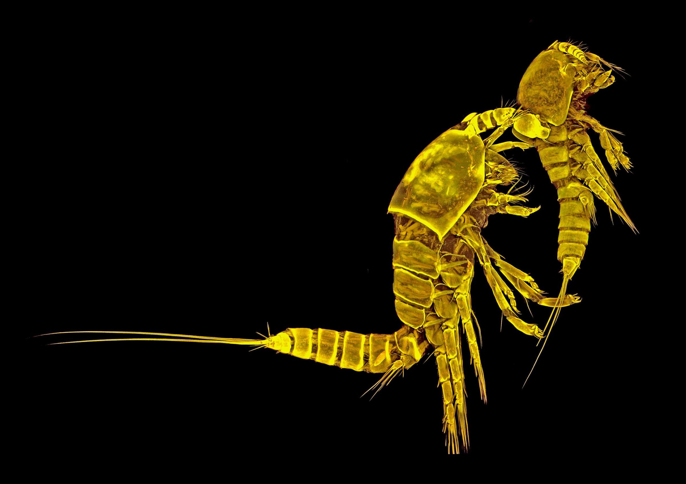
Tigriopus californicus (copepod), couple, lateral view
A Tigriopus californicus (copepod) at 10x magnification.Dr. Terue Kihara
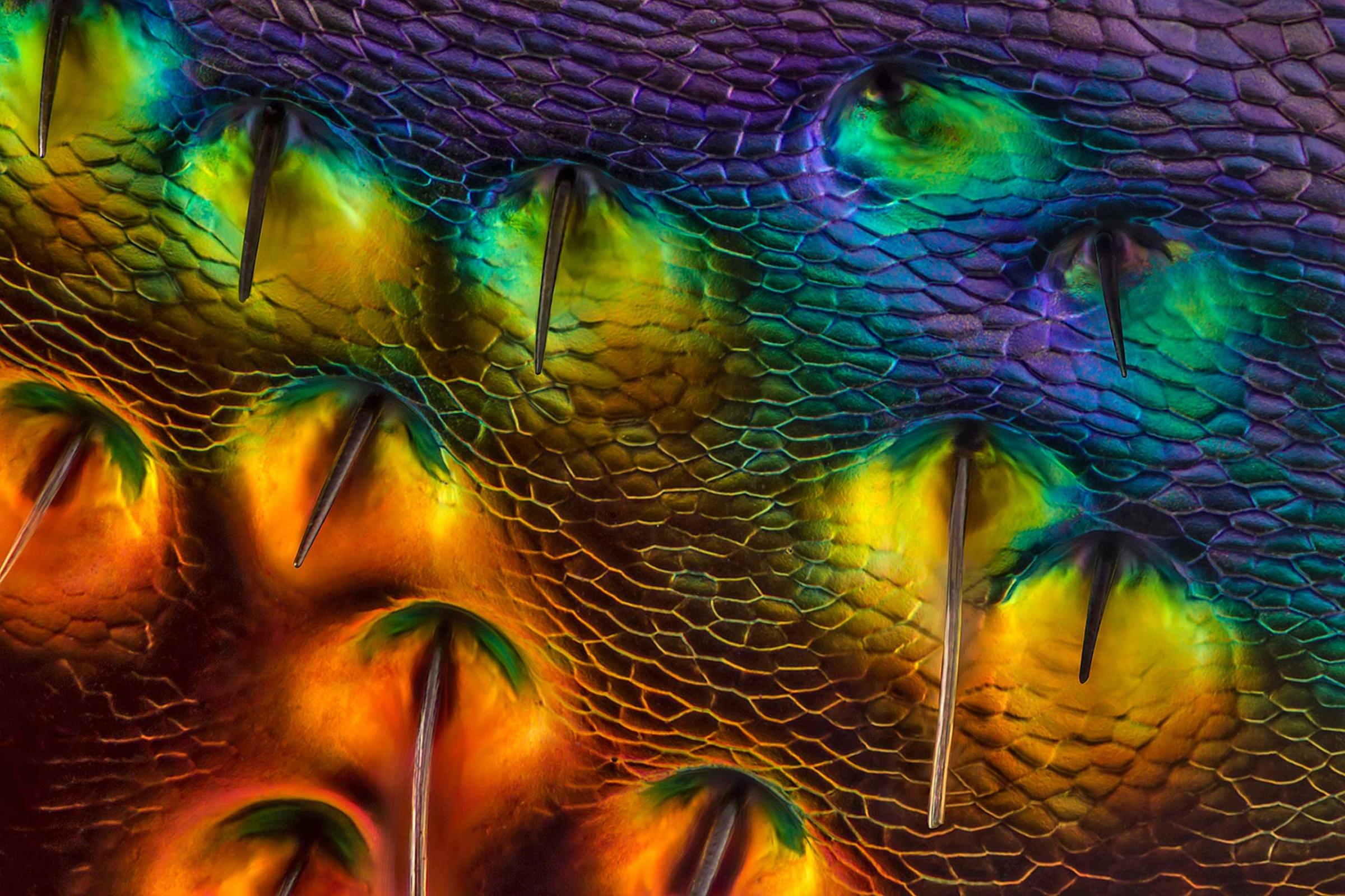
Chrysochroa buqueti (jewel beetle) carapace, near eye
A Chrysochroa buqueti (jewel beetle) carapace, near eye at 450x magnification.Charles Krebs
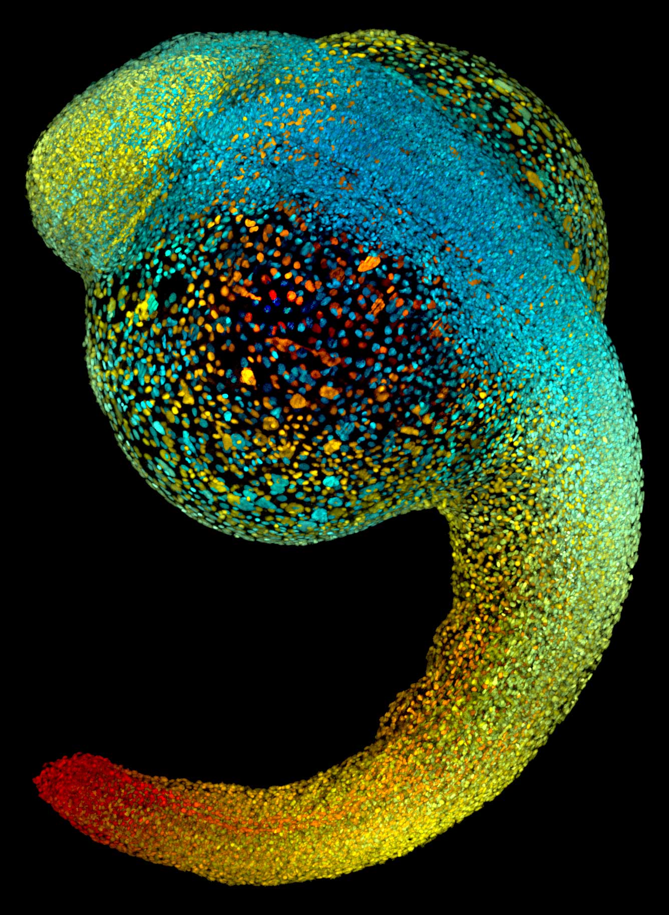
Live zebrafish embryo at 22 hours post-fertilization
Live zebrafish embryo at 22 hours post-fertilization.Dr. Philipp Keller
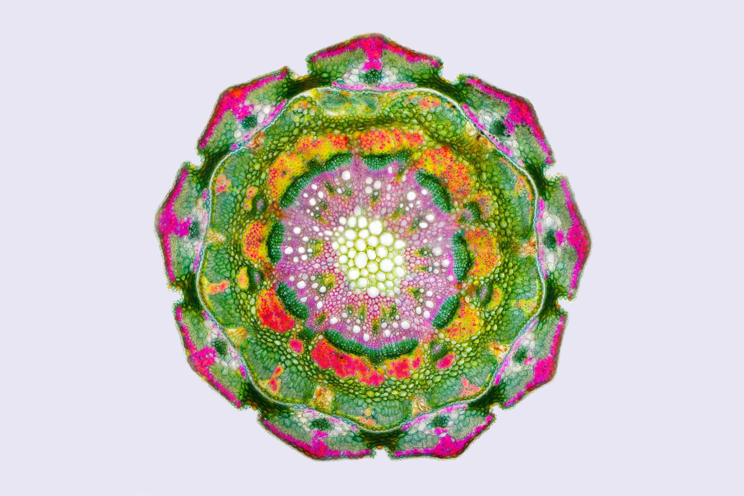
Casuarina equisetifolia (beach oak) twigs with scale leaves, transverse section
A Casuarina equisetifolia (beach oak) twigs at 125x magnification.Anatoly Mikhaltsov
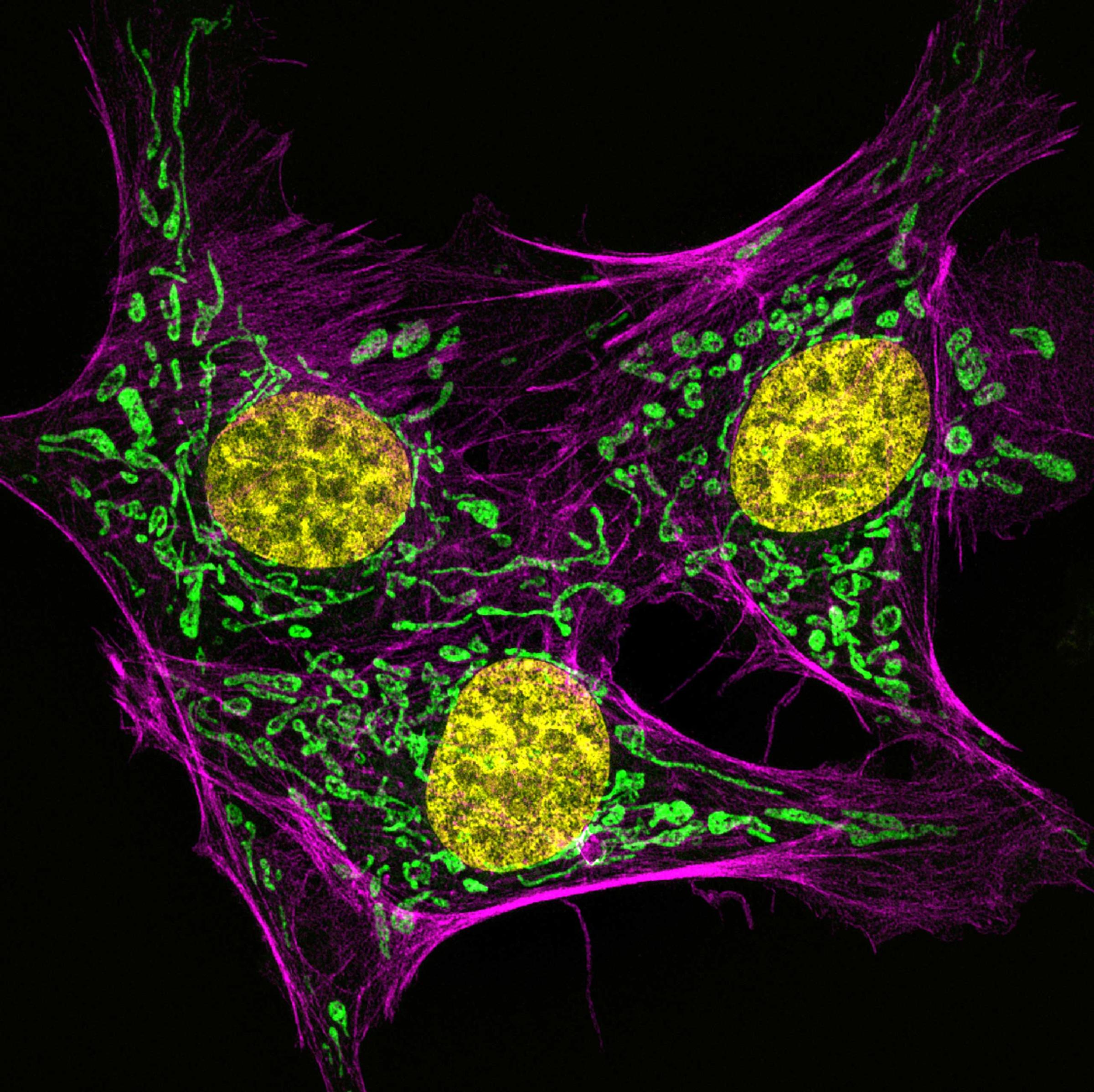
Bovine pulmonary artery endothelial cells stained for actin (pink), mitochondria (green) and DNA (yellow
Bovine pulmonary artery endothelial cells.Dr. Muthugapatti K. Kandasamy
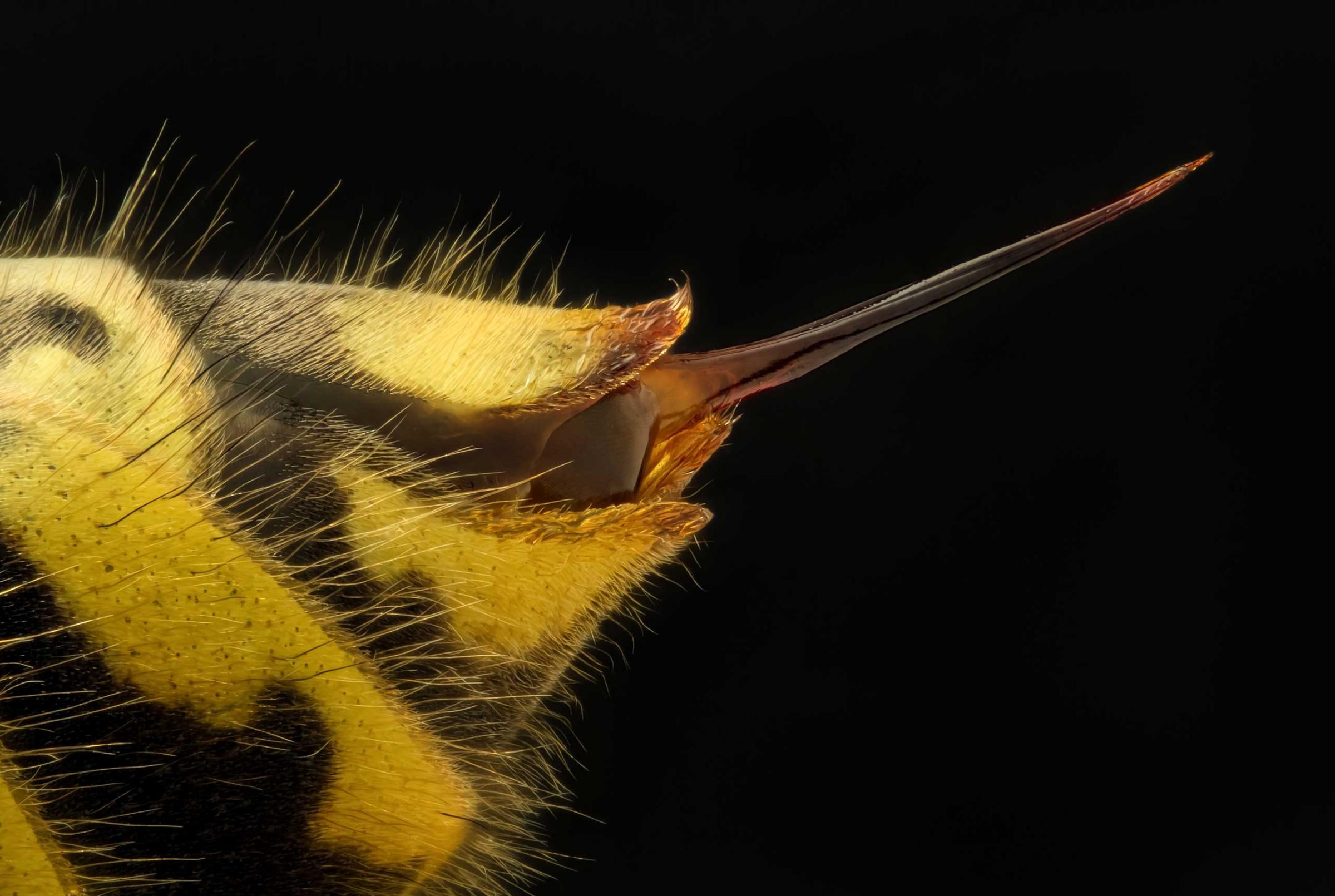
A Vespula vulgaris (common wasp) stinger at 5x magnification.Geir Drange
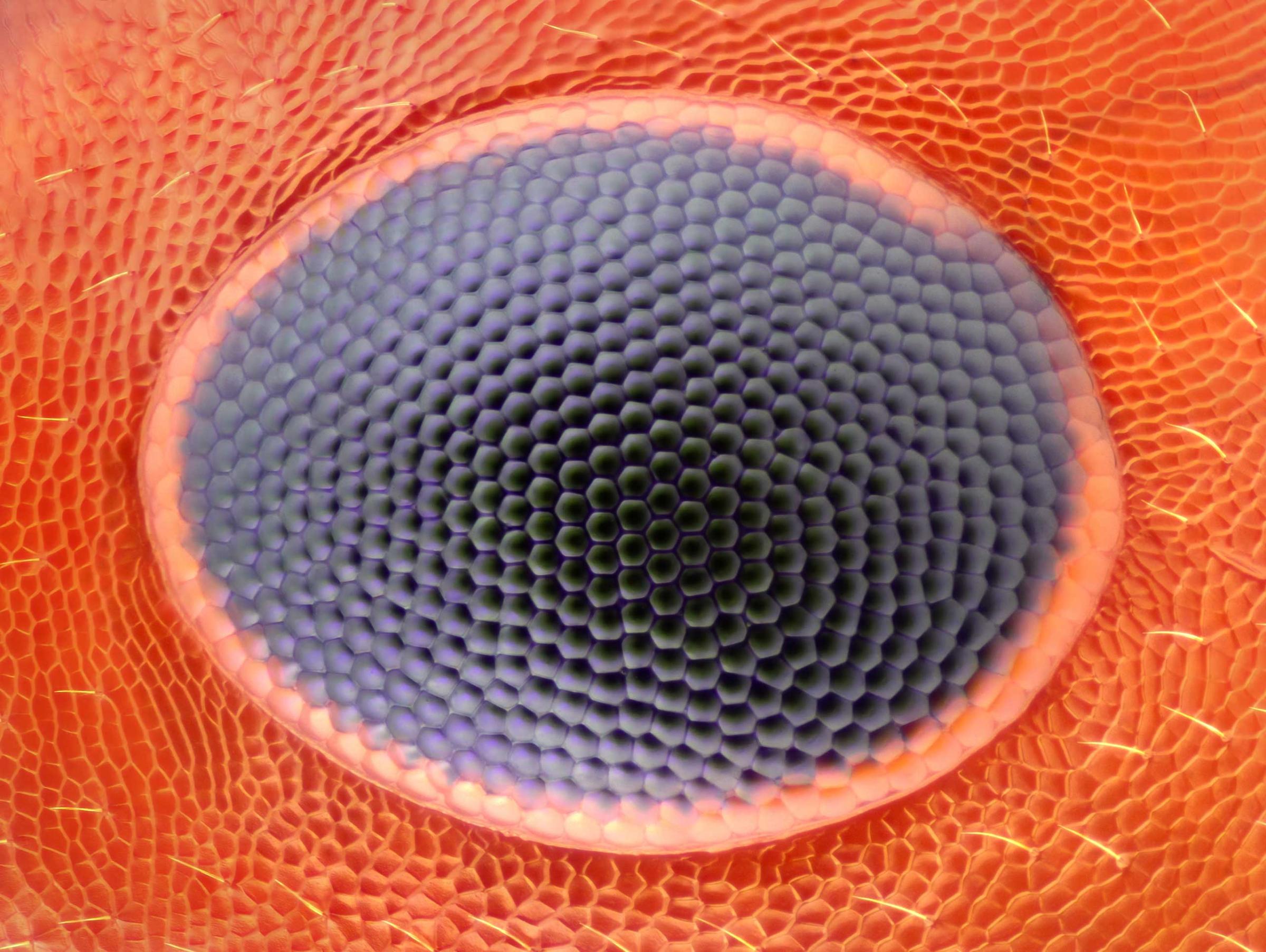
Ant Eye
An ant eye at 20x magnification.Noah Fram-Schwartz
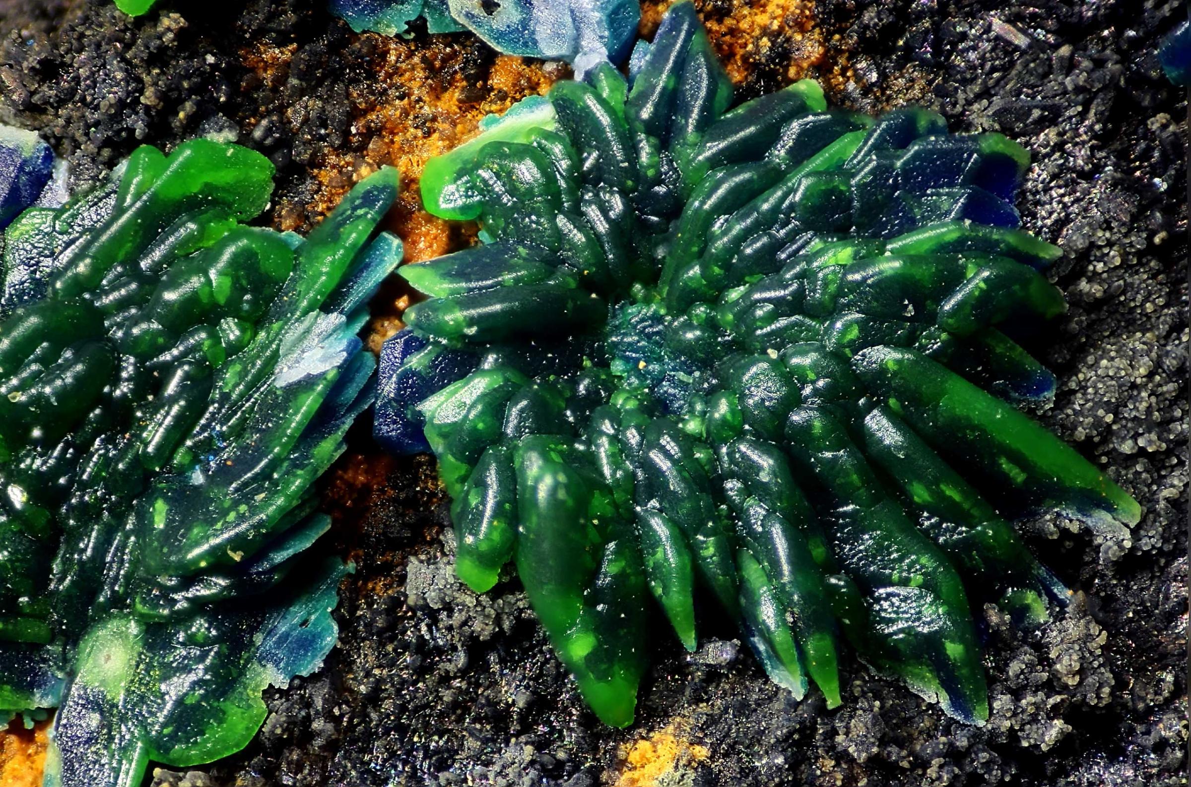
Conichalcite pseudomorph after azurite
Conichalcite pseudomorph after azurite at 6x magnification.Honorio Cócera-La Parra
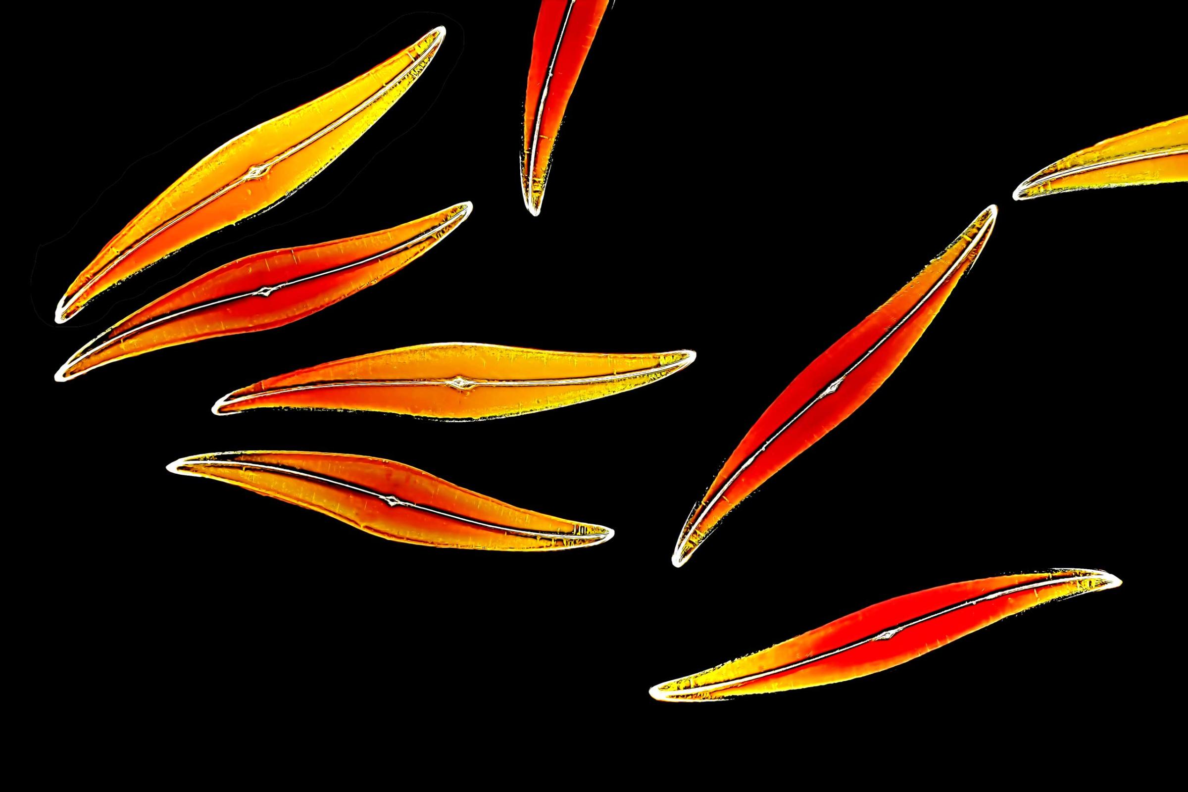
Pleurosigma angulatum (diatoms
Pleurosigma angulatum (diatoms) at 100x magnification.Christian Gautier
Lilium anther, second division tetrads
Lilium anther, second division tetrads at 500x magnification.Raymond Sloss
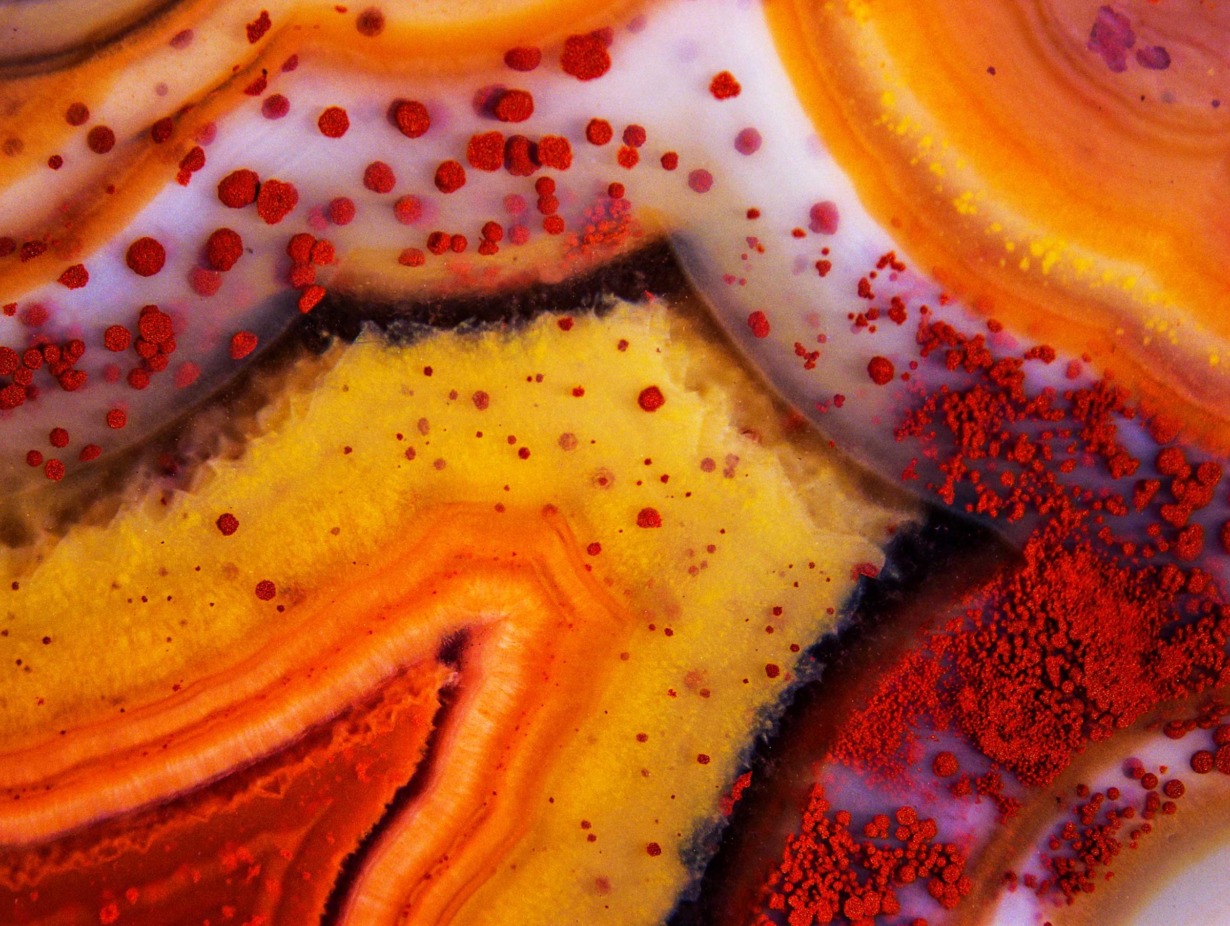
Montana Dryhead agate, unpolished
Montana Dryhead agate at 50x magnification.Douglas L. Moore
Tripolycyanamide crystal
Tripolycyanamide crystal at 100x magnification.Yanping Wang
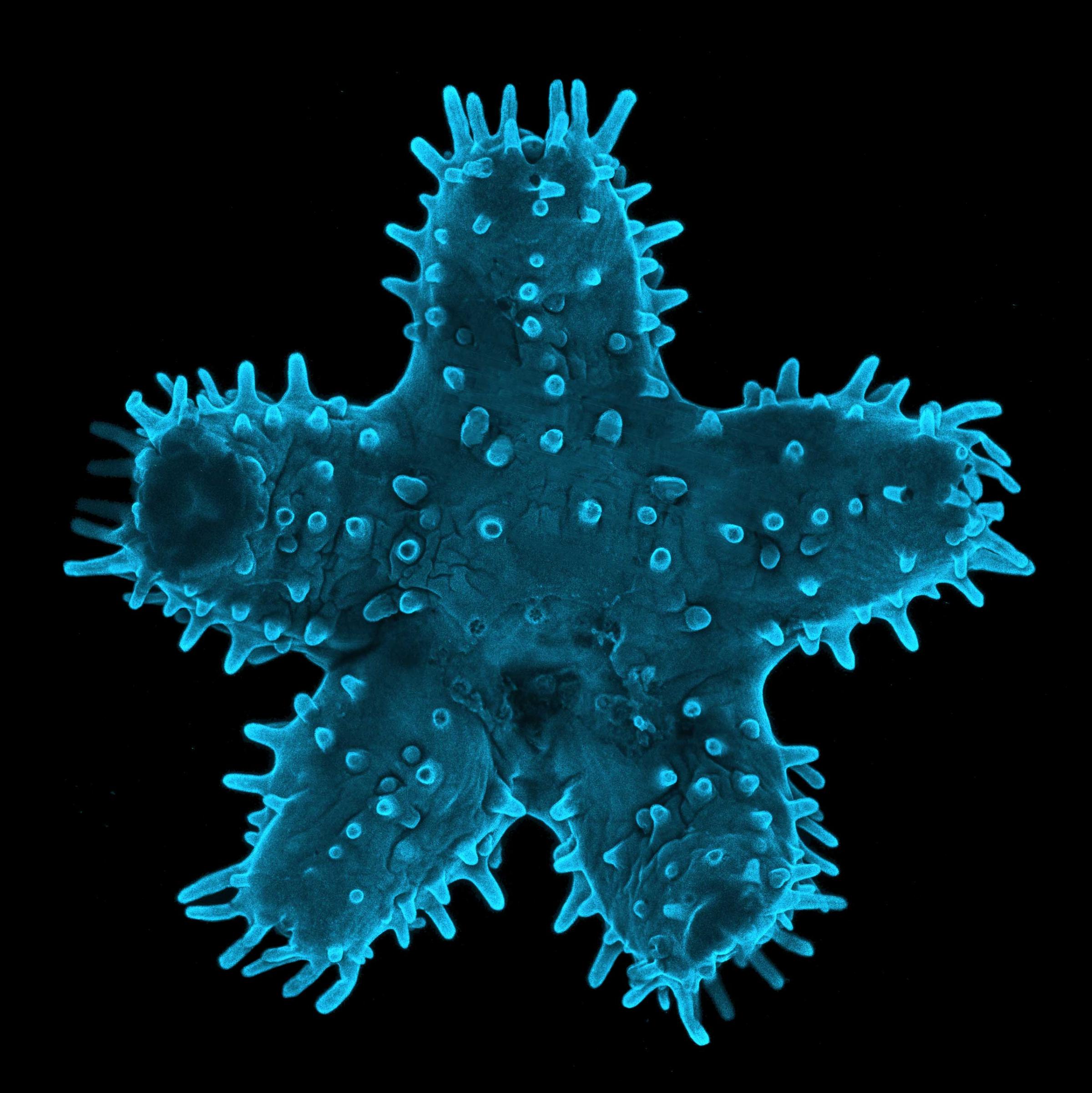
Young Starfish
A young starfish at 5x magnification.Steven Wilbert
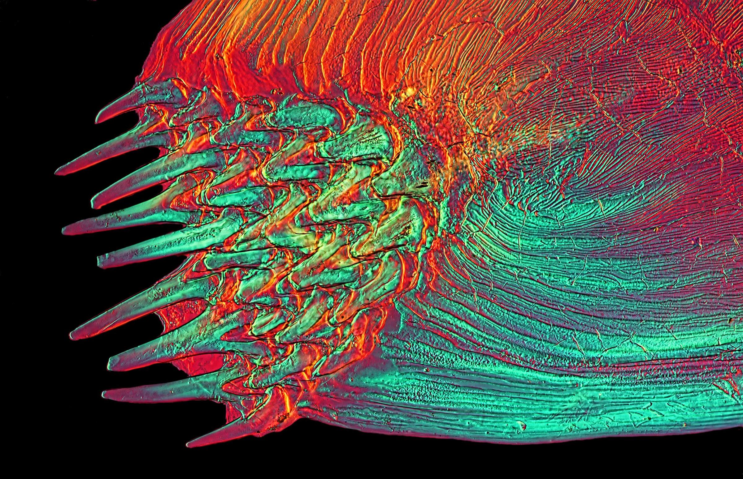
A Solea sp. (fish) at 25x magnification.David Linstead
Pleurotaenium ovatum
Pleurotaenium ovatum (micro algae) at 40x magnification.Rogelio Moreno Gill
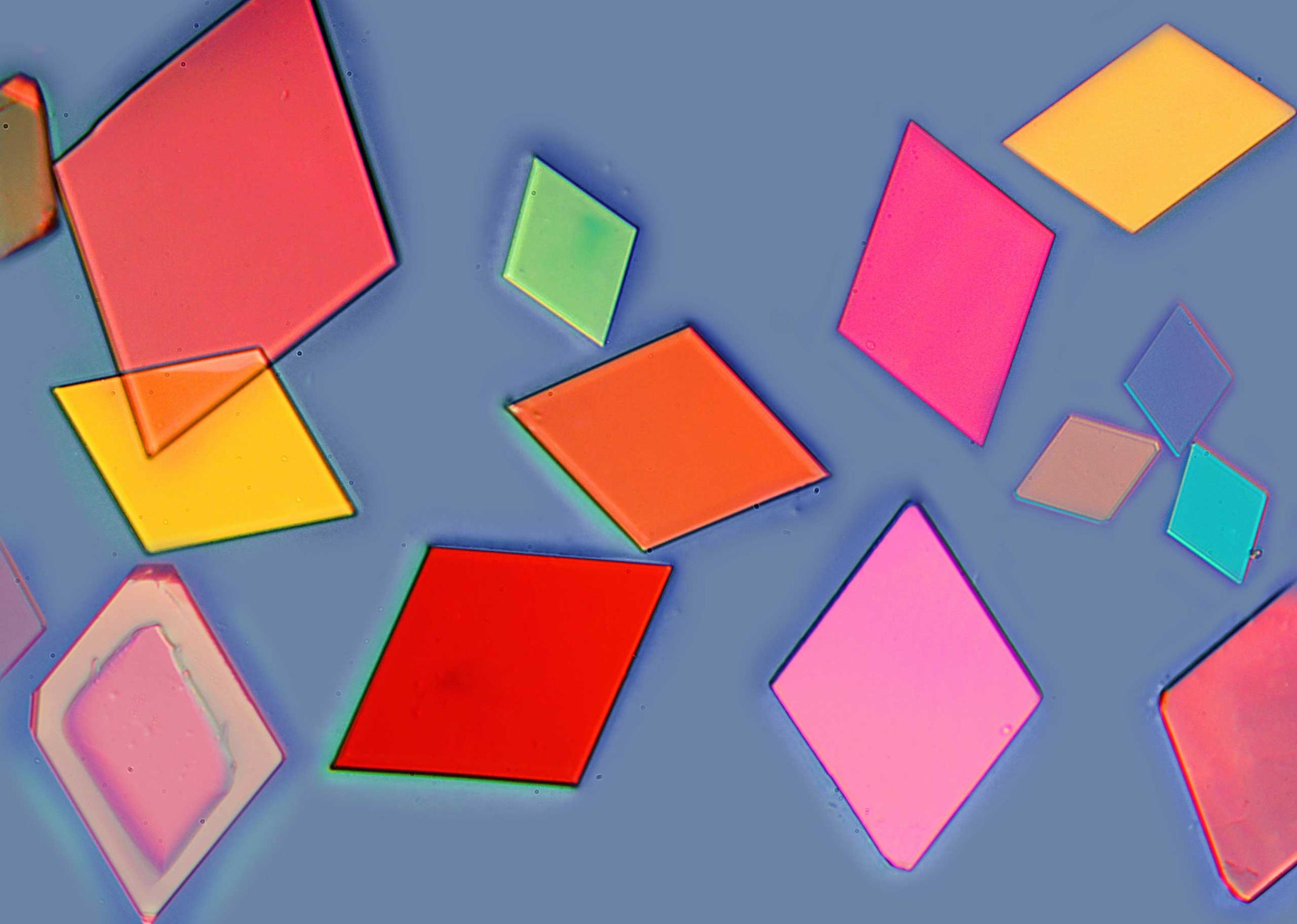
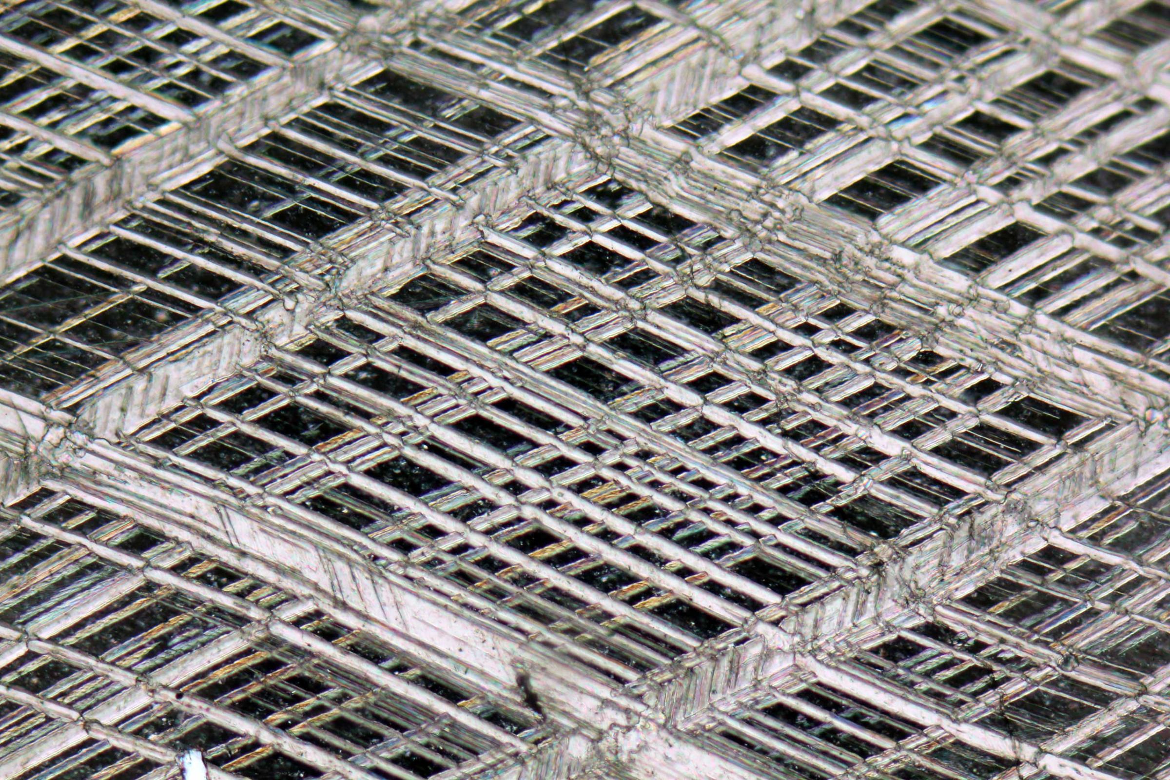
A Magnesium chloride and potassium alum mixture at 25x magnification.Chao Zhang
A rhombohedral cleavage in calcite crystal at 10x magnification.Alessandro Da Mommio
Autofluorescence in marine algae
Marine algae at 40x magnification. Waldo Nell
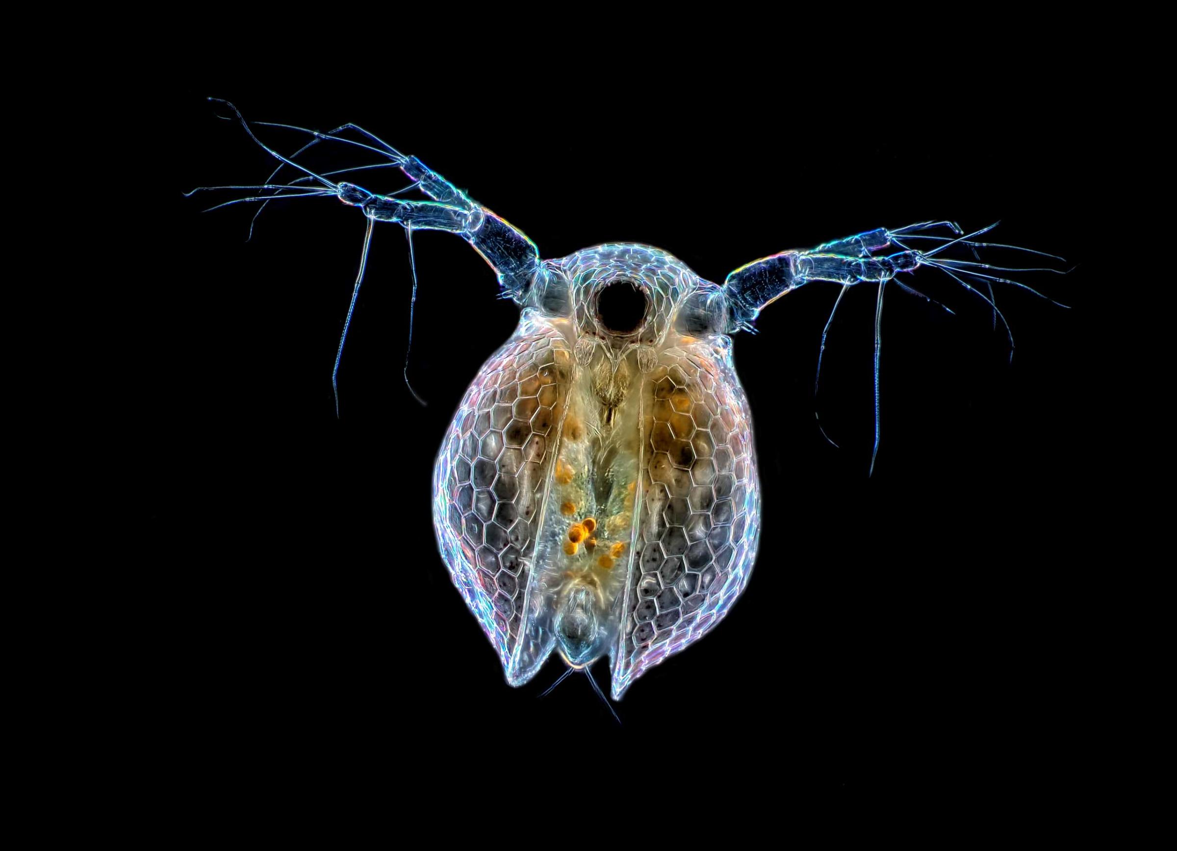
A Ceriodaphnia sp. (water flea) at 20x magnification.Rogelio Moreno Gill
Snowflake
A snowflake at 8x magnification.Michael Peres
Shipworm Lyrodus pedicellatus, a wood-boring mussel
A Shipworm Lyrodus pedicellatus and a wood-boring mussel at 20x magnification.Andrea Wurzinger-Mayer
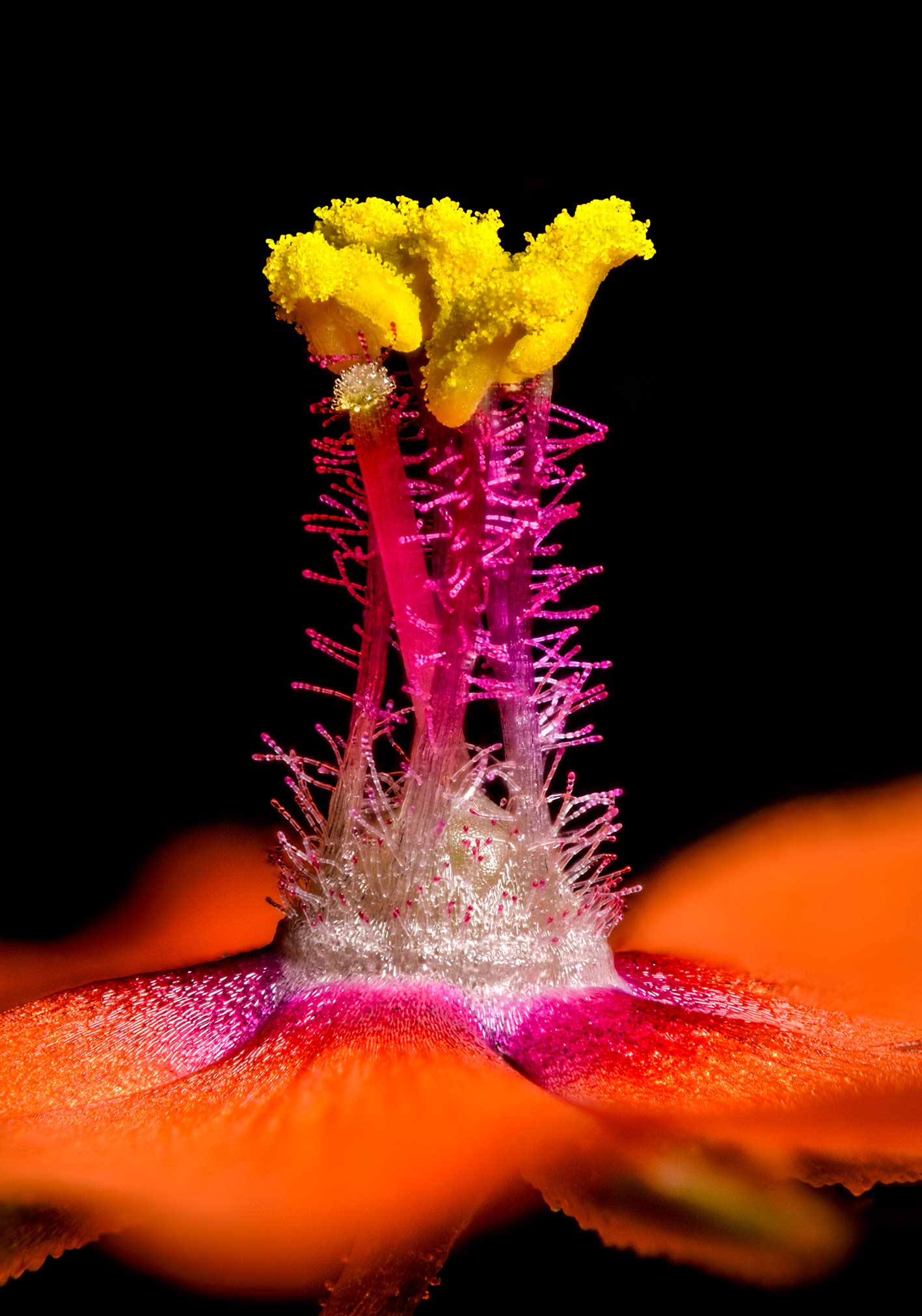
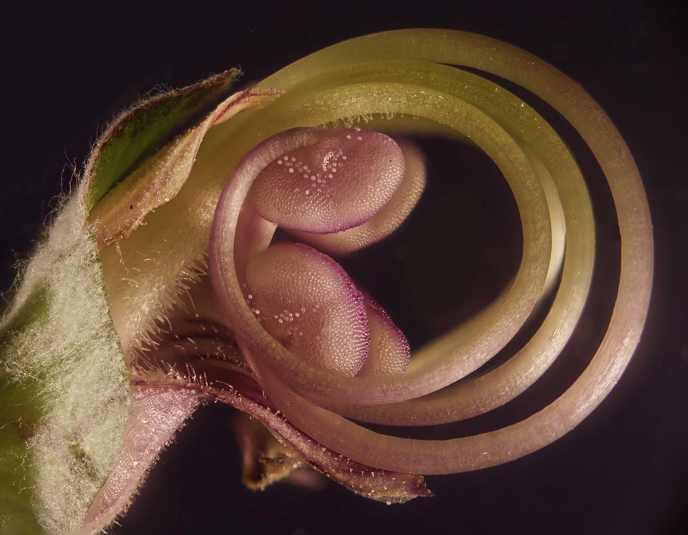
Anagallis arvensis (scarlet pimpernel)
Anagallis arvensis (scarlet pimpernel) at 80x magnification. Jens H. Petersen
Flower embryo
A flower embryo at 40x magnification.Samuel Silberman
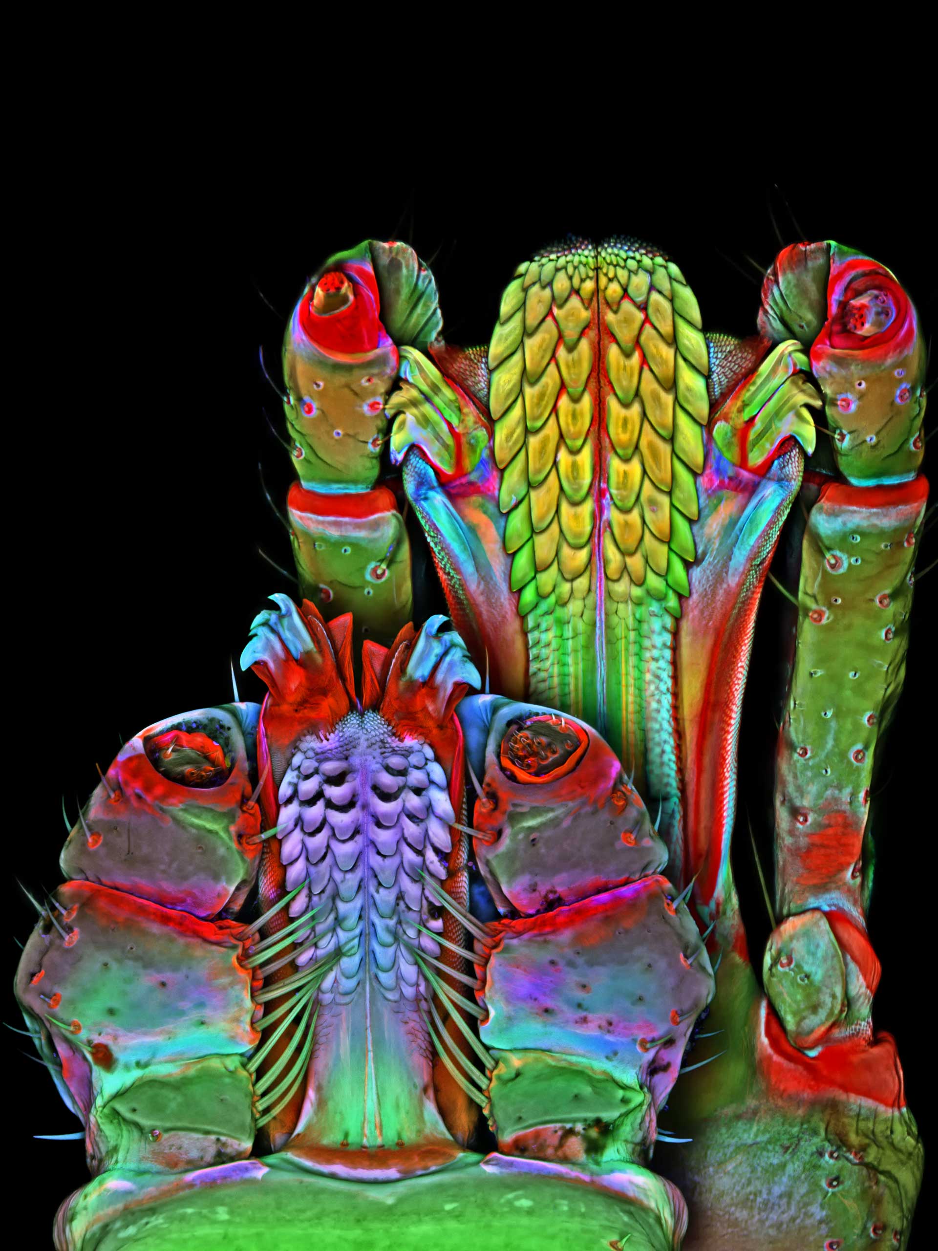
Underside of the Brown dog tick and Lonestar tick mouthparts
The underside of a Brown dog tick and Lonestar tick at 100x magnification.Dr. Igor Robert Siwanowicz
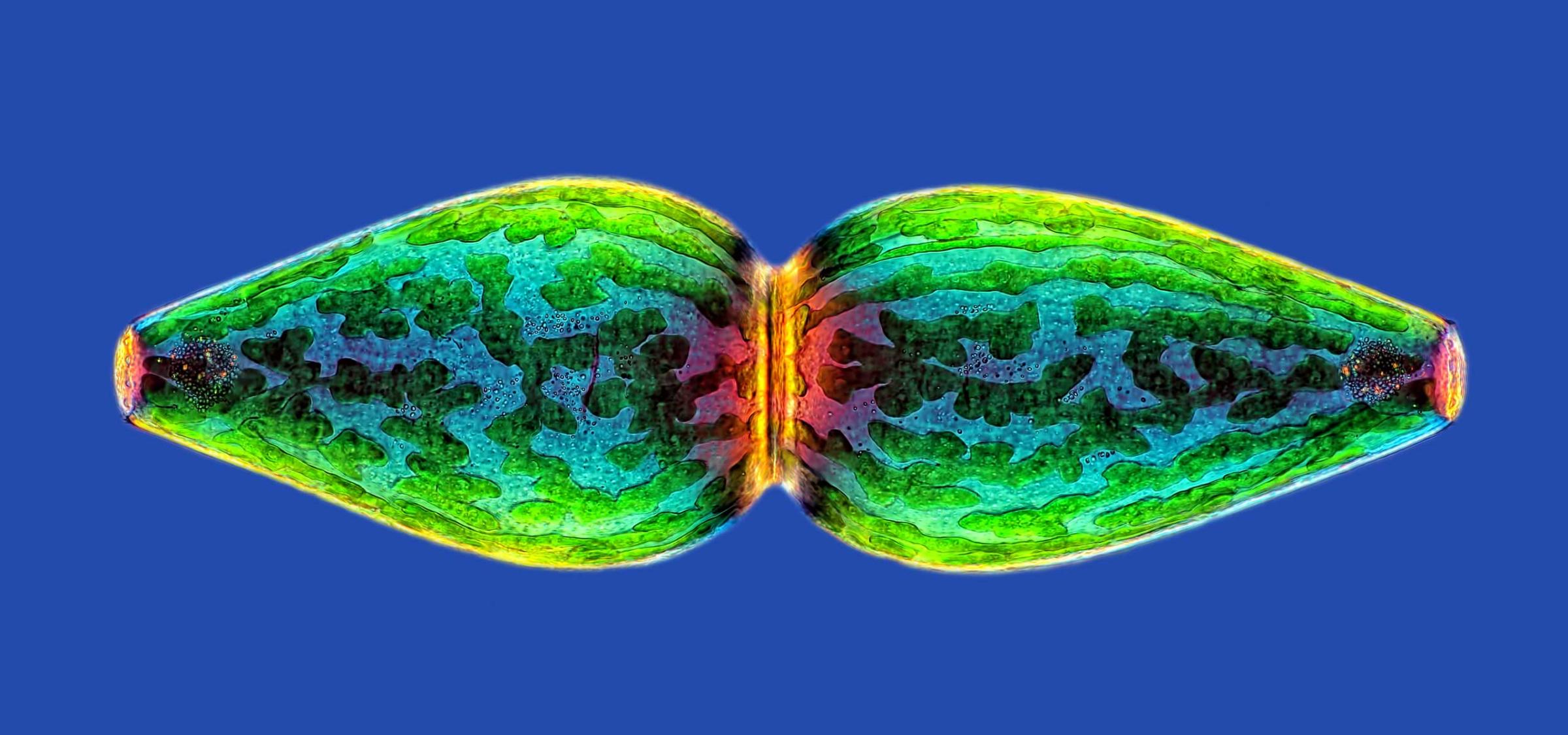
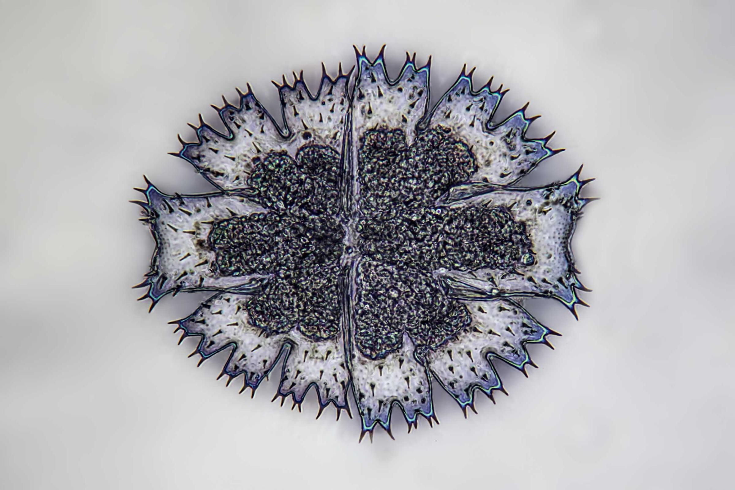
Living Micrasterias in contrast Interphako at 100 magnification.Frank Fox
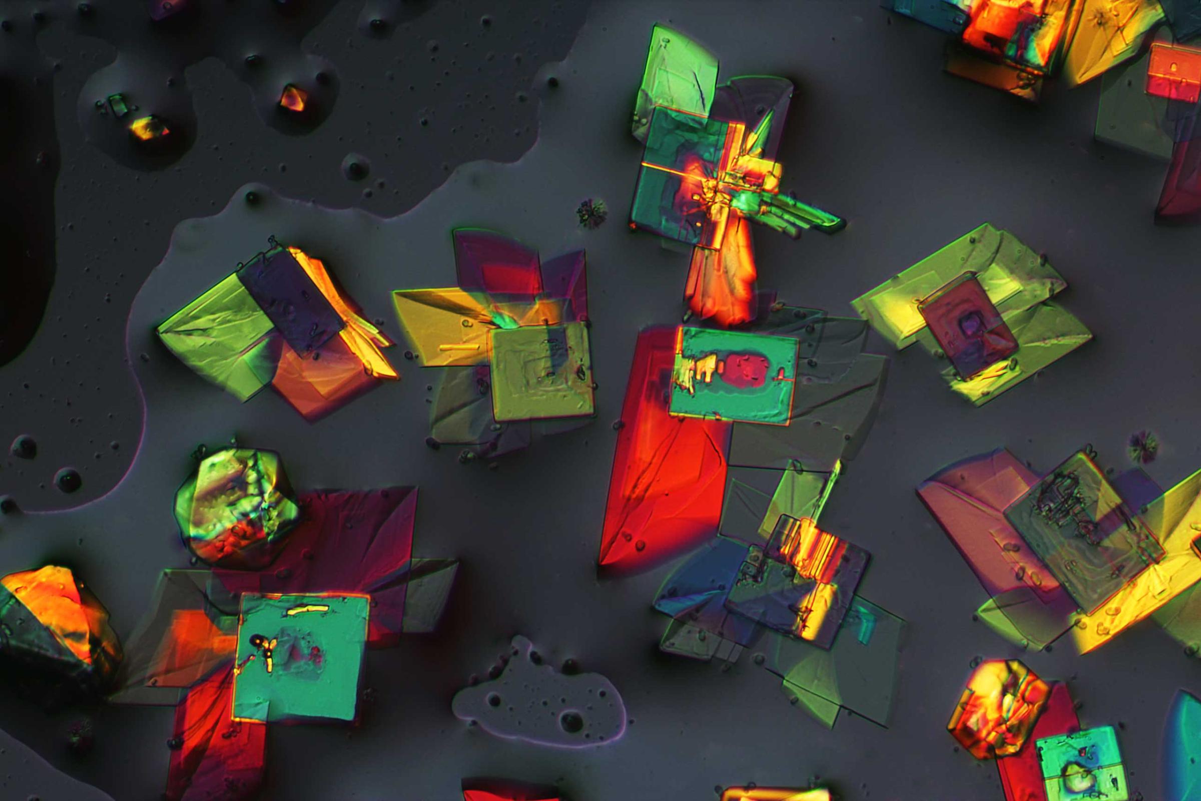
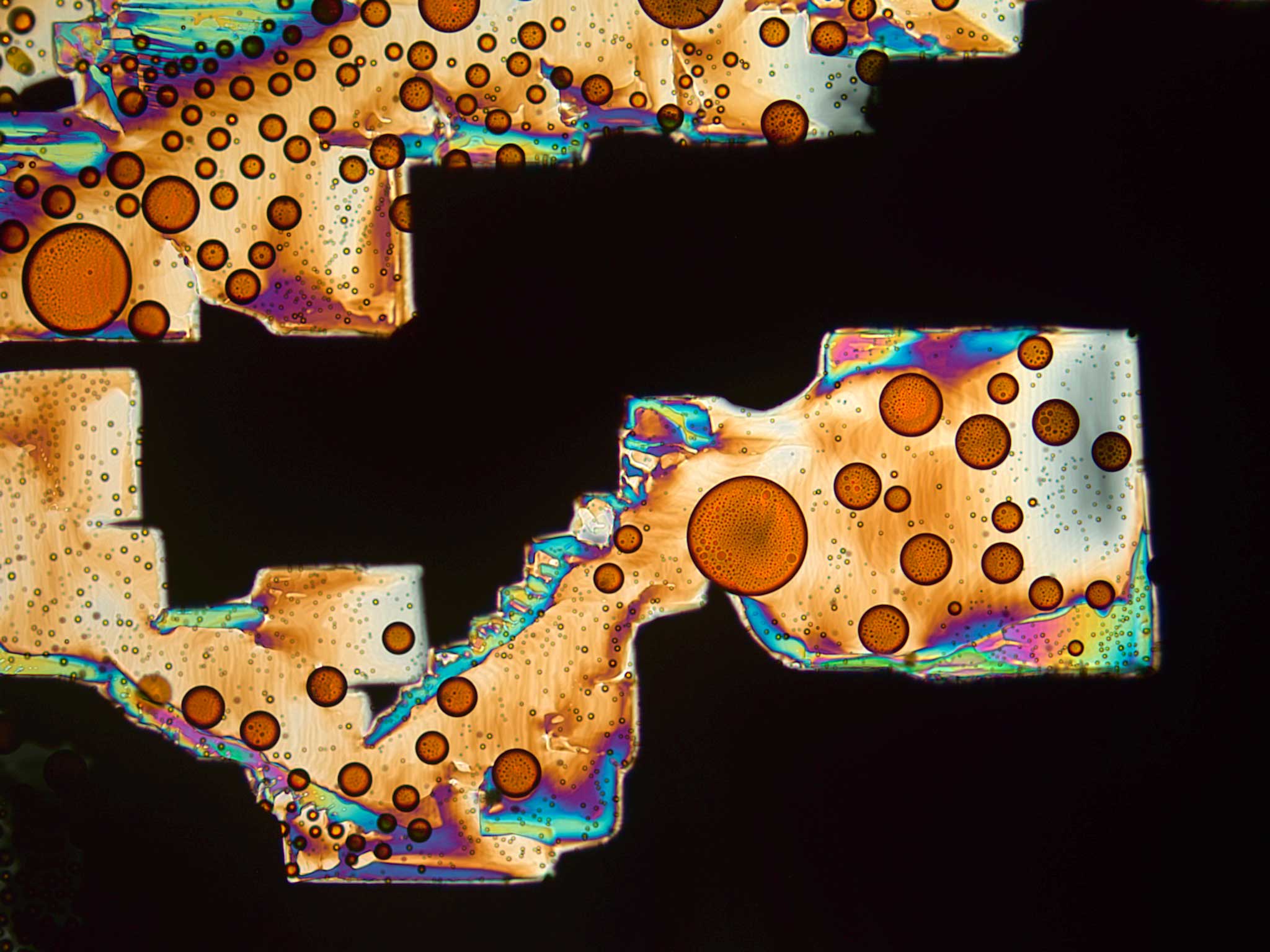
Recrystallized bismuth iodide under crossed polars
Recrystallized bismuth iodide at 100x magnification.Sebastian Blaise Sparenga
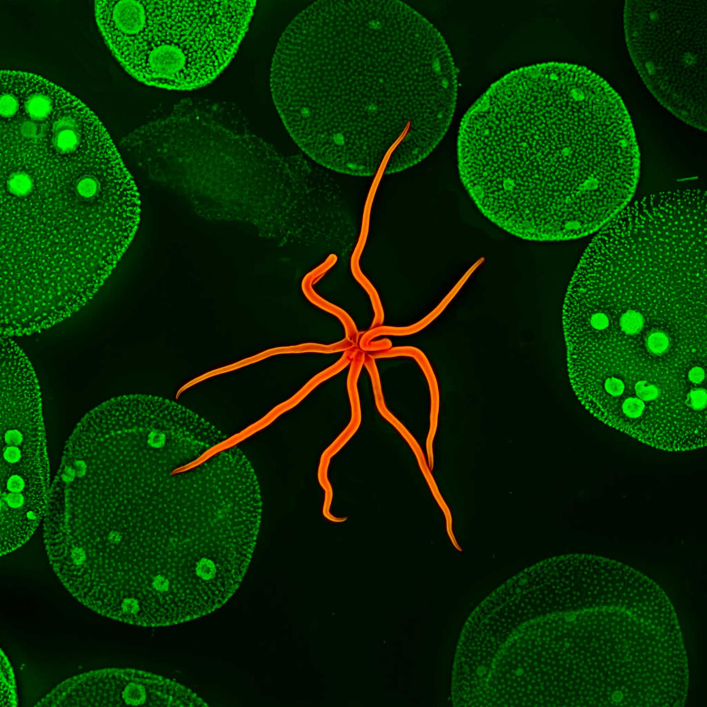
Hydra and Volvox
A Hydra and Volvox at 10x magnification.Steven Wilbert
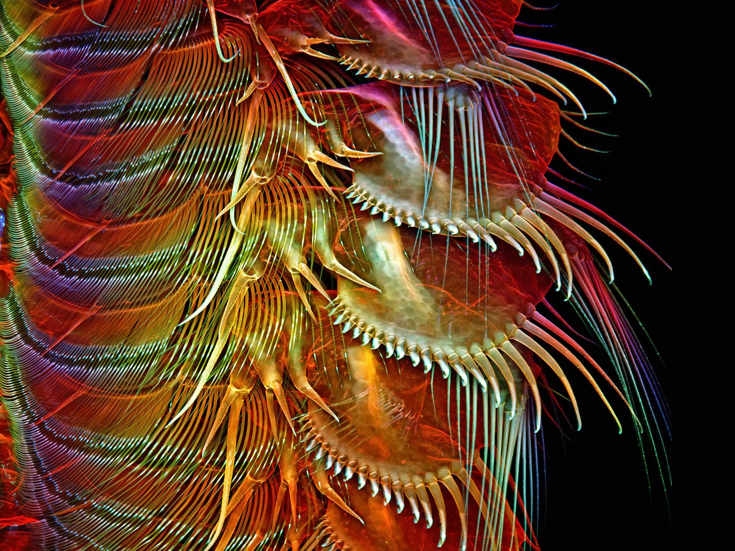
Appendages of a common brine shrimp
Appendages of a common brine shrimp at 100x magnification.Dr. Igor Robert Siwanowicz

Read next: What’s the World’s Deadliest Creature?

Download TIME’s mobile app for iOS to have your world explained wherever you go

More Must-Reads From TIME

Contact us at letters@time.com