Stalking a Silent Killer: Photos From the Early Fight Against Cancer
Stalking a Silent Killer: Photos From the Early Fight Against Cancer
3 minute read
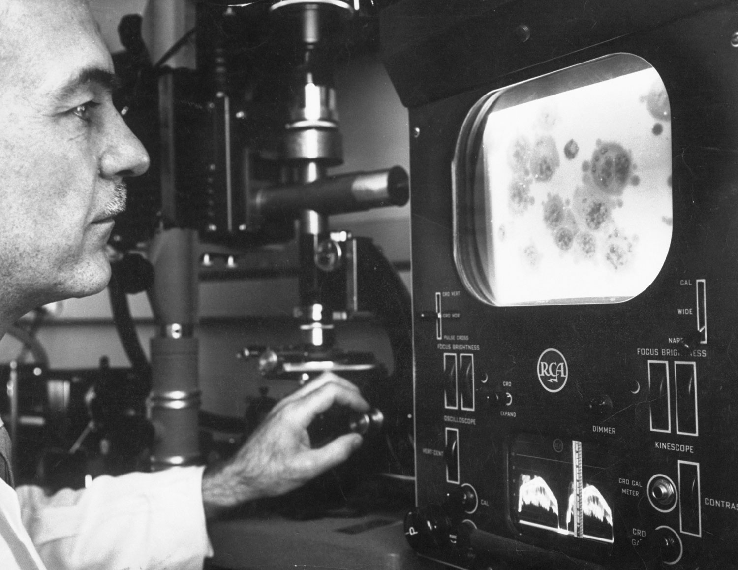
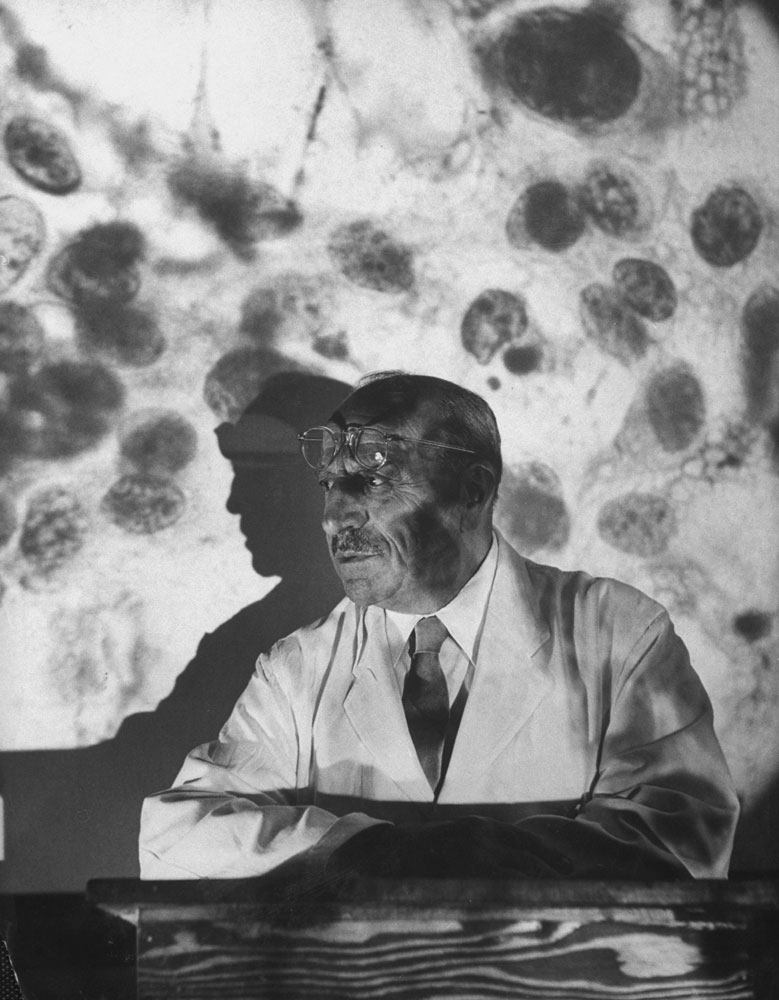
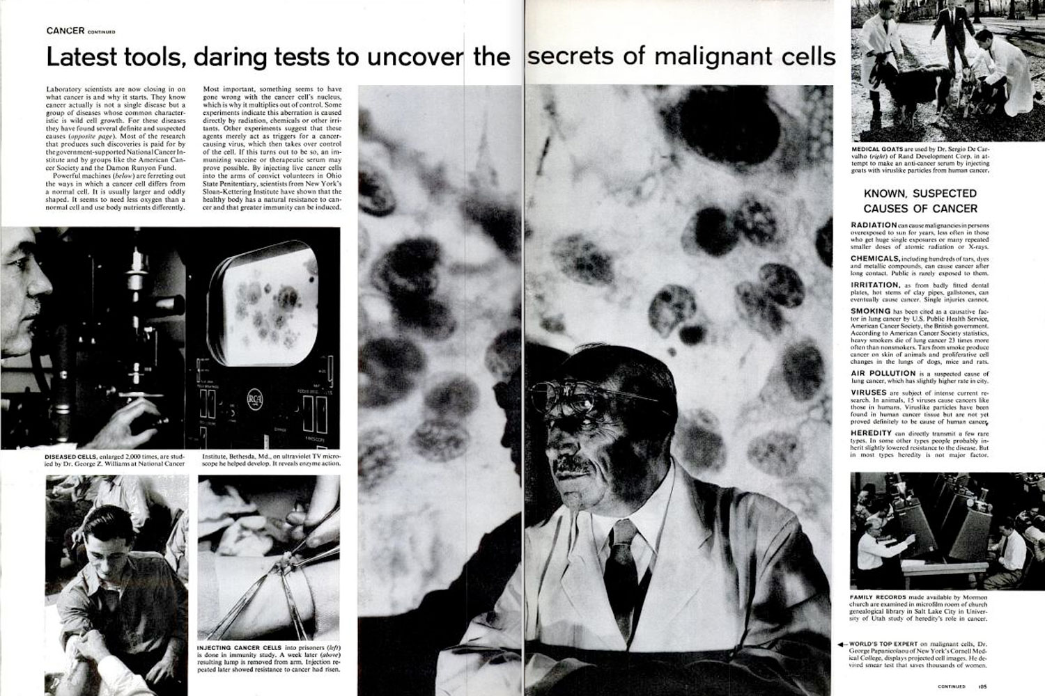
Caption from LIFE. "Diseased cells, enlarged 2,000 times, are studied by Dr. George Z. Williams at National Cancer institute, Bethesda, Md., on ultraviolet TV microscope he helped develop. It reveals enzyme action."Andreas Feininger—Time & Life Pictures/Getty Images
Angelina Jolie’s bombshell announcement that she underwent a preventive double mastectomy in hopes of minimizing her risk of breast cancer still has, understandably, an awful lot of people talking. Some commentators have suggested that Jolie’s action and, more importantly, the powerful, public way that she let the world know of her decision — on her own schedule, in her own words — could end up saving lives, if only because so many women who might have put off testing for years are now avidly searching for all the information they can find on breast cancer.
Of course, guarded optimism (and open pessimism) in the face of cancer is hardly new. Or rather, the last half century or so has seen truly extraordinary strides in treatment of myriad types of the disease. Sixty-five years ago, in May 1958, LIFE magazine published a cover story that considered an arsenal of new medical, technological and chemical approaches to diagnosis and treatment — approaches, the magazine suggested, that might do away with some cancers within the lifetime of those then fighting them.
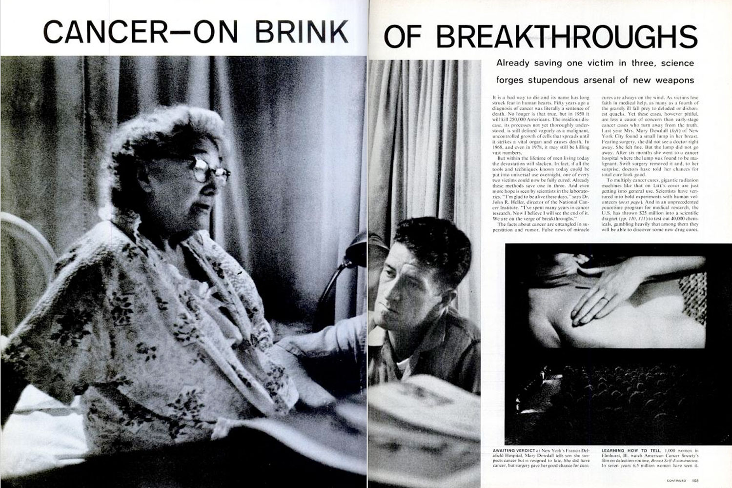
The article, titled titled, “Cancer — on Brink of Breakthroughs,” put it this way to LIFE’s millions of readers:
It is a bad way to die and its name has long struck fear in human hearts. Fifty years ago a diagnosis of cancer was literally a sentence of death. No longer is that true, but in 1958 it will kill 250,000 Americans. [That number has more than doubled in the past 65 years. Last year, the American Cancer Society released a report stating that “in 2012, about 577,190 Americans are expected to die of cancer, more than 1,500 people a day. Cancer is the second most common cause of death in the US, exceeded only by heart disease, accounting for nearly 1 of every 4 deaths. — Ed.]
But within the lifetime of men living today the devastation will slacken. In act, if all the tools and techniques known today could be put into universal use overnight, one of every two victims could now be fully cured. Already these methods save one in three.
The facts about cancer are tangled in superstition and rumor. False news of miracle cures are always on the wind. As victims lose faith in medical help, as many as a fourth of the gravely ill fall prey to deluded or dishonest quacks.
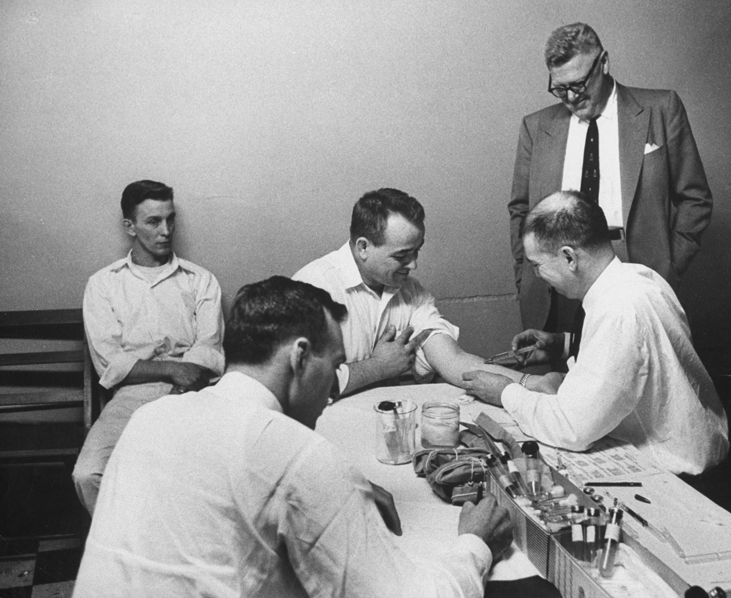
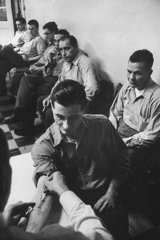
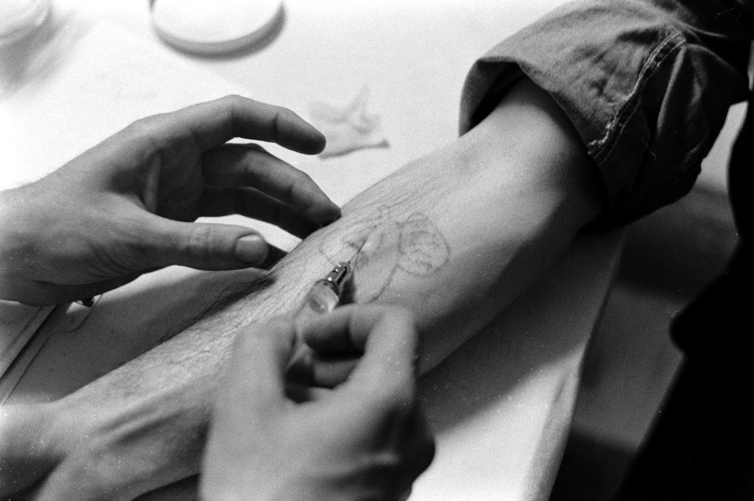
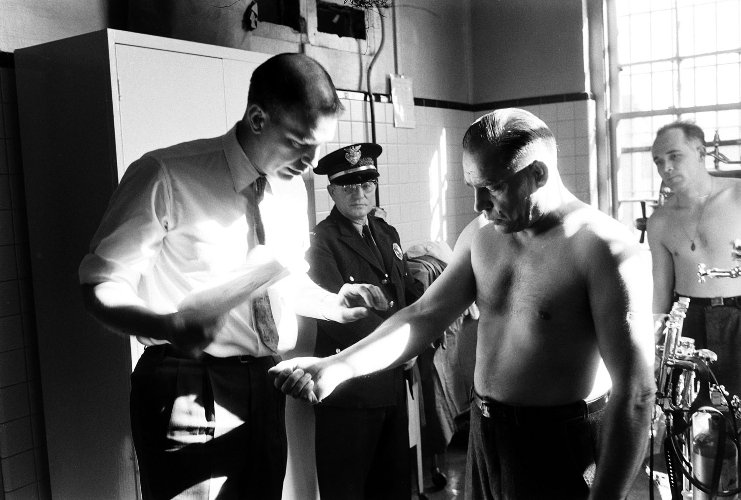
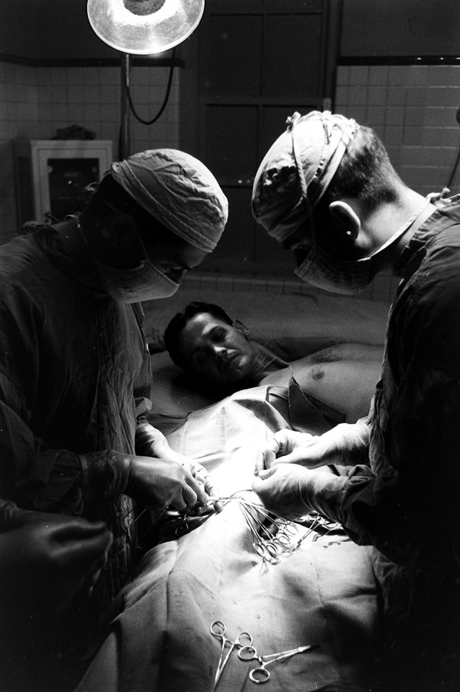
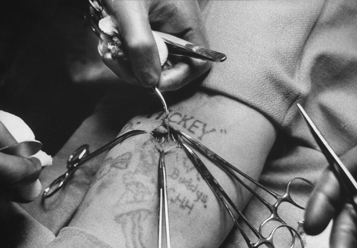
Some of the photos in this gallery, meanwhile, will no doubt surprise and even shock contemporary readers. Prisoners injected with cancerous cells in order to test patient’s resistance to some cancers after exposure? Obviously, testing (of drugs, surgeries, exposure to pathogens, etc.) on both animals and on humans has always been a central component of the practice of medicine. But rarely does one see as graphic a representation of the way such tests were conducted in the middle part of the last century as in some of the pictures in this gallery.
Liz Ronk, who edited this gallery, is the Photo Editor for LIFE.com. Follow her on Twitter @lizabethronk.
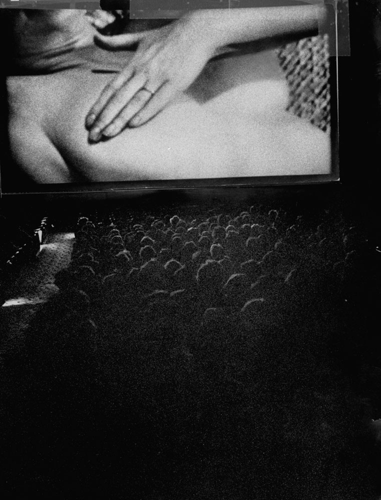
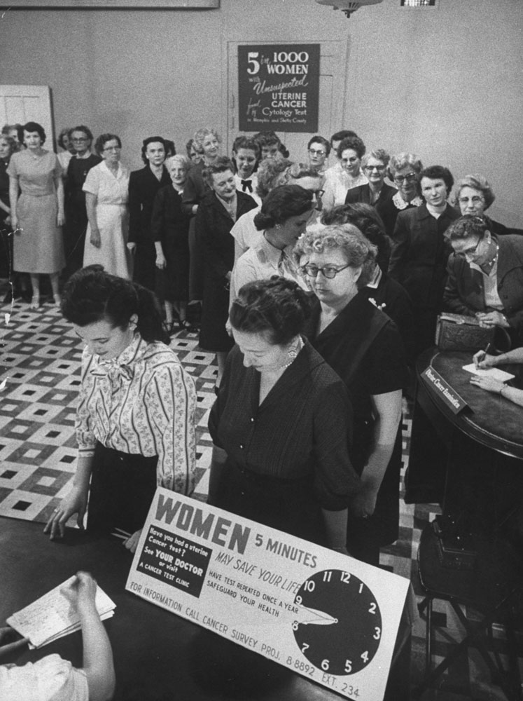
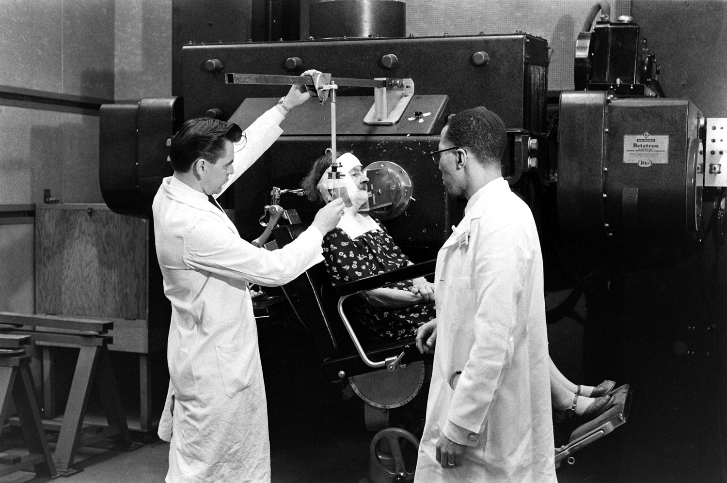
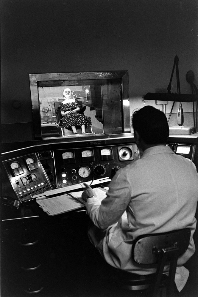
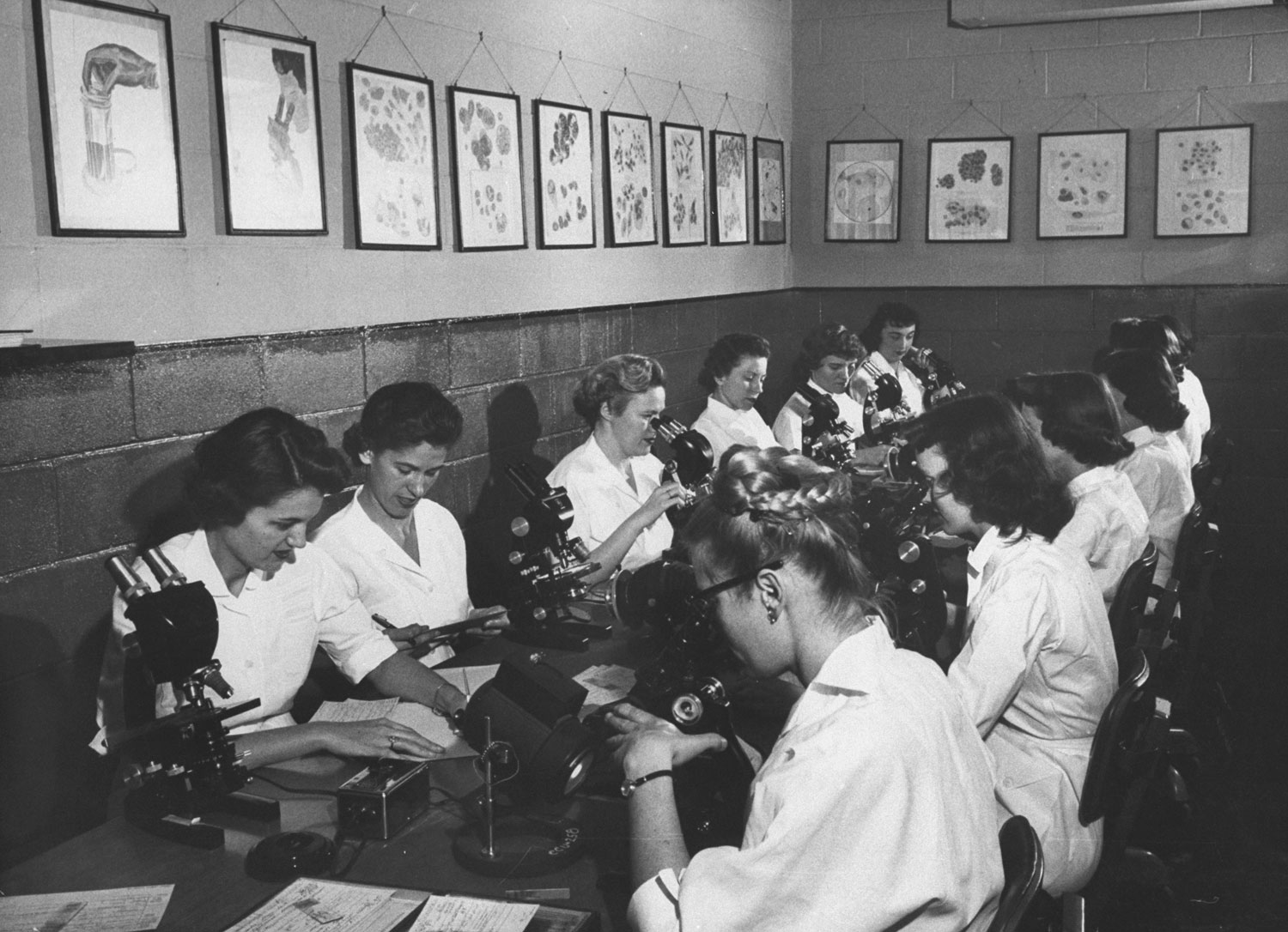
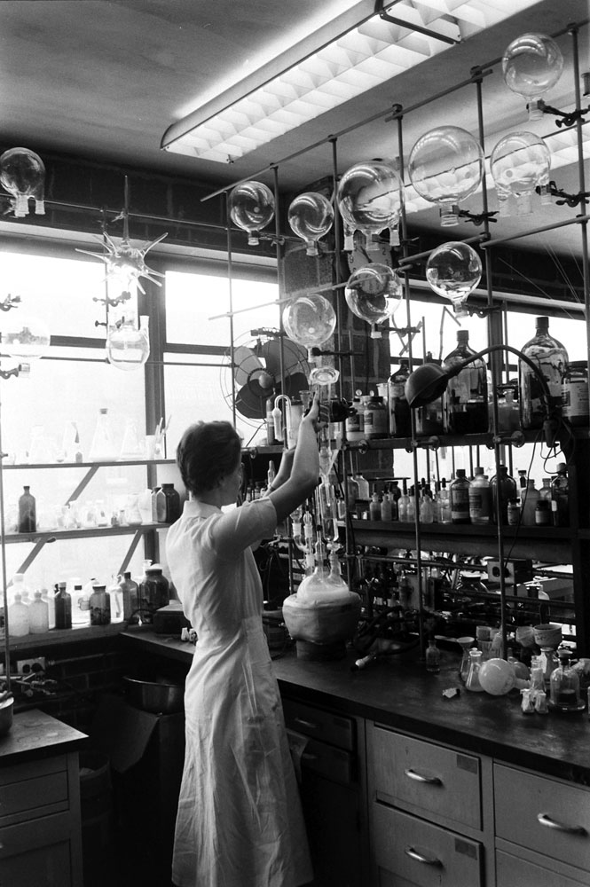
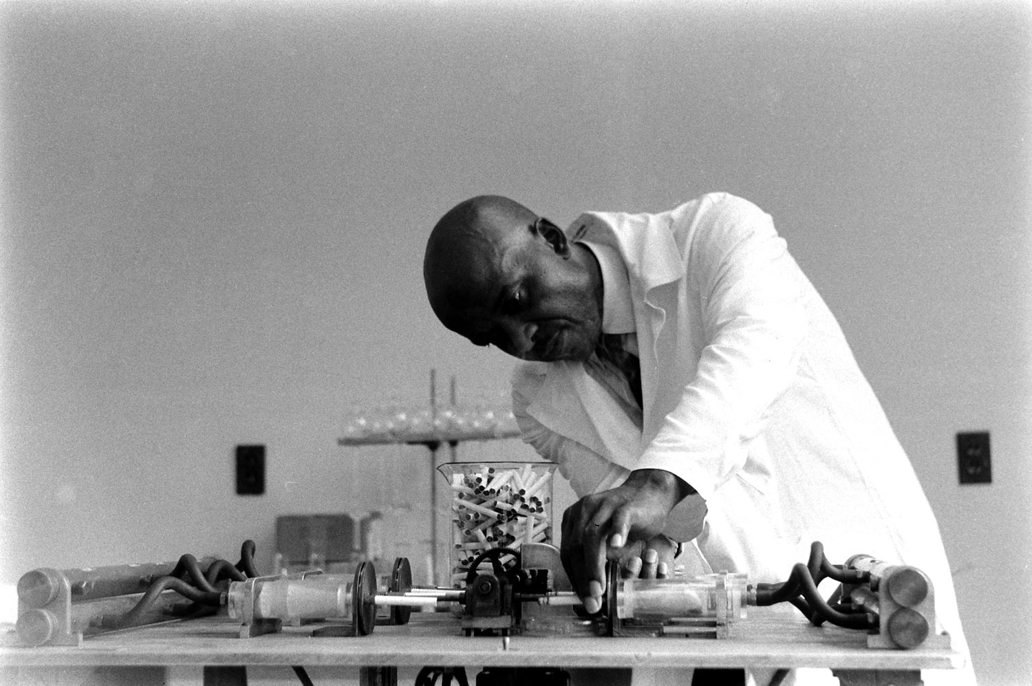
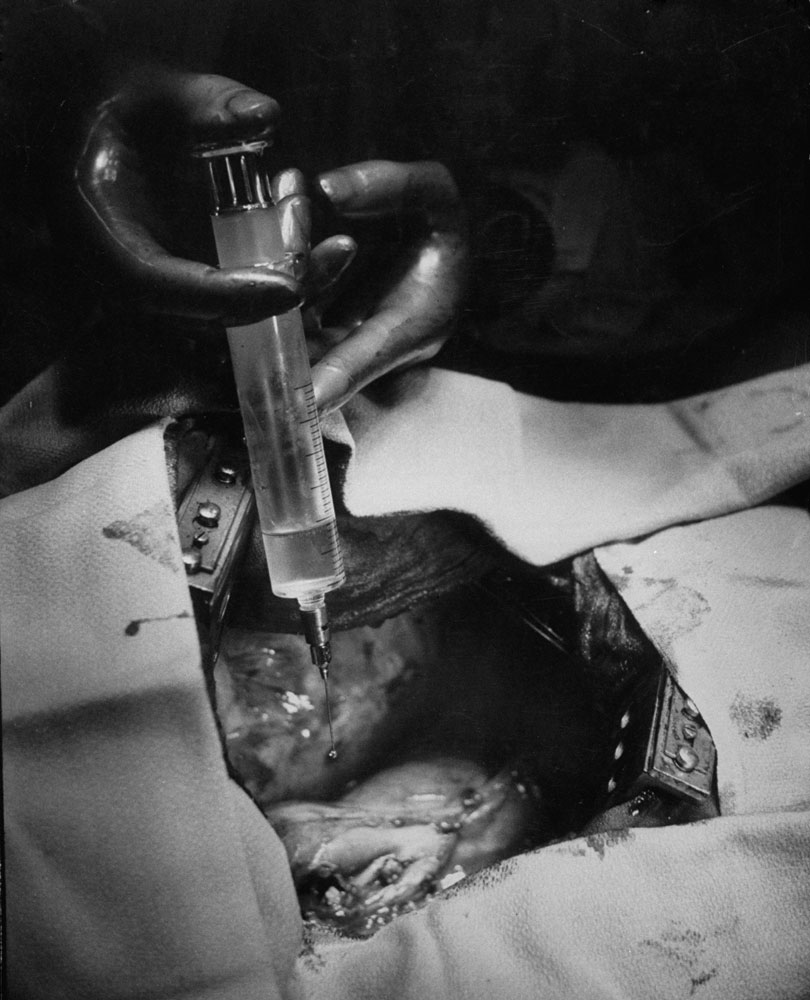
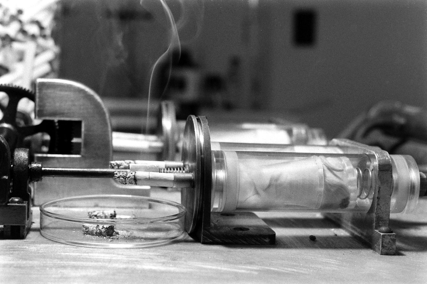
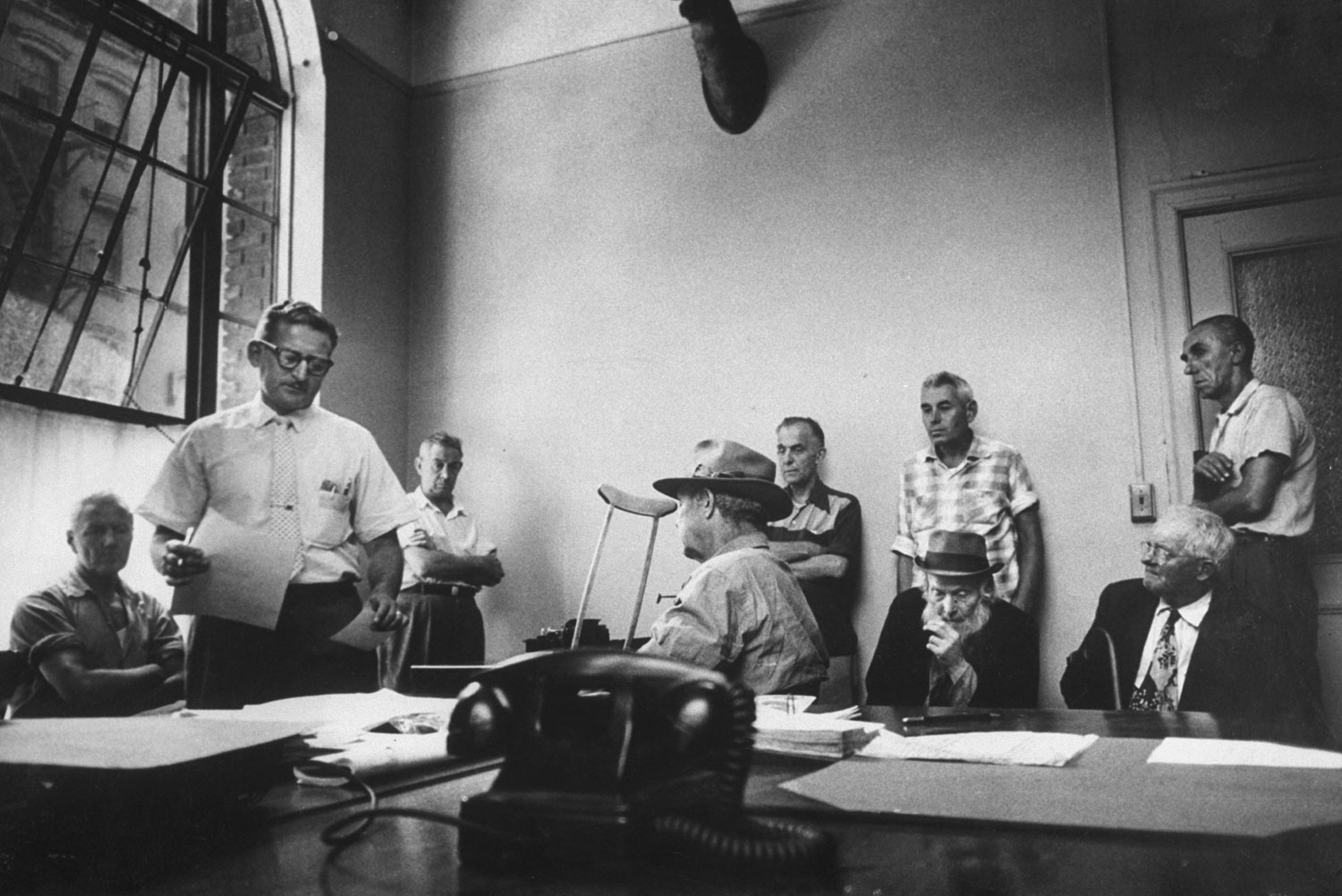
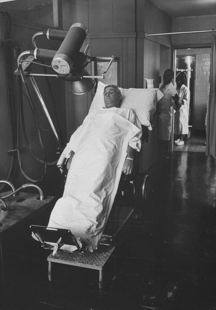
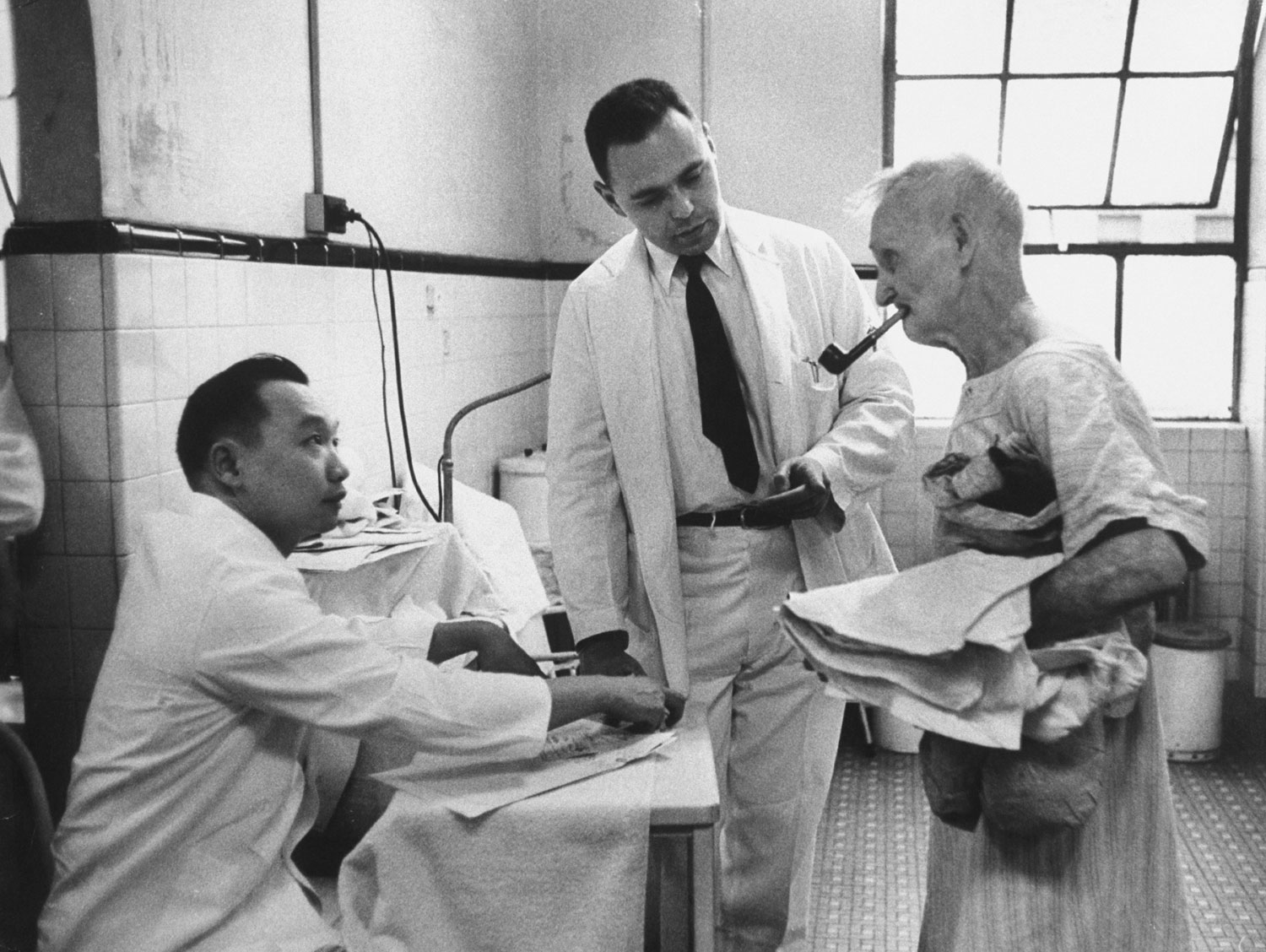
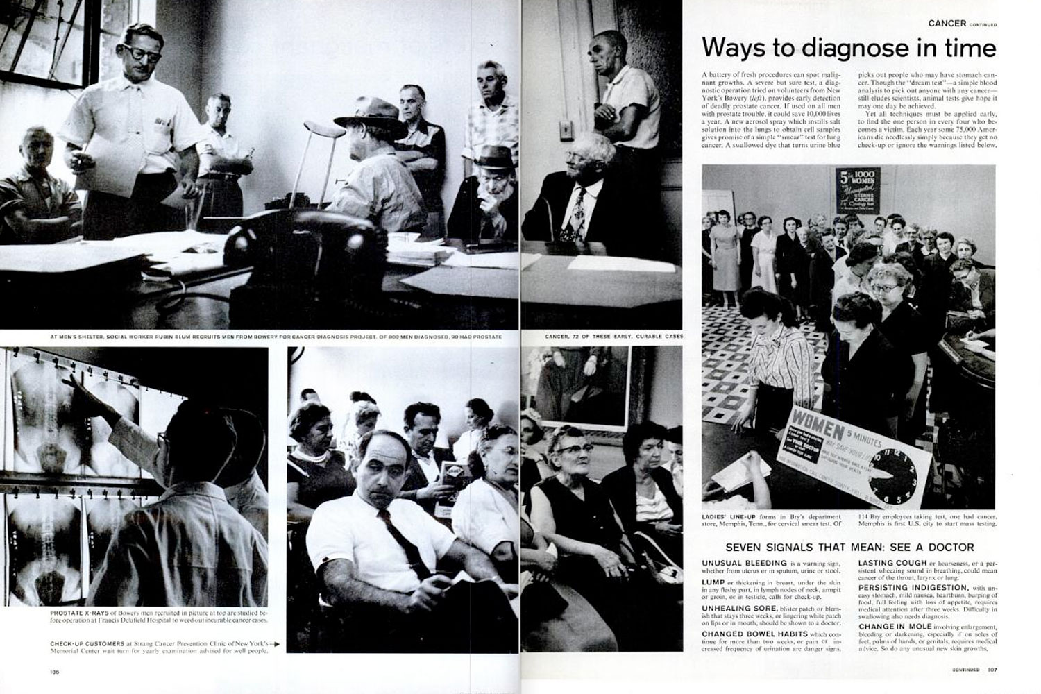
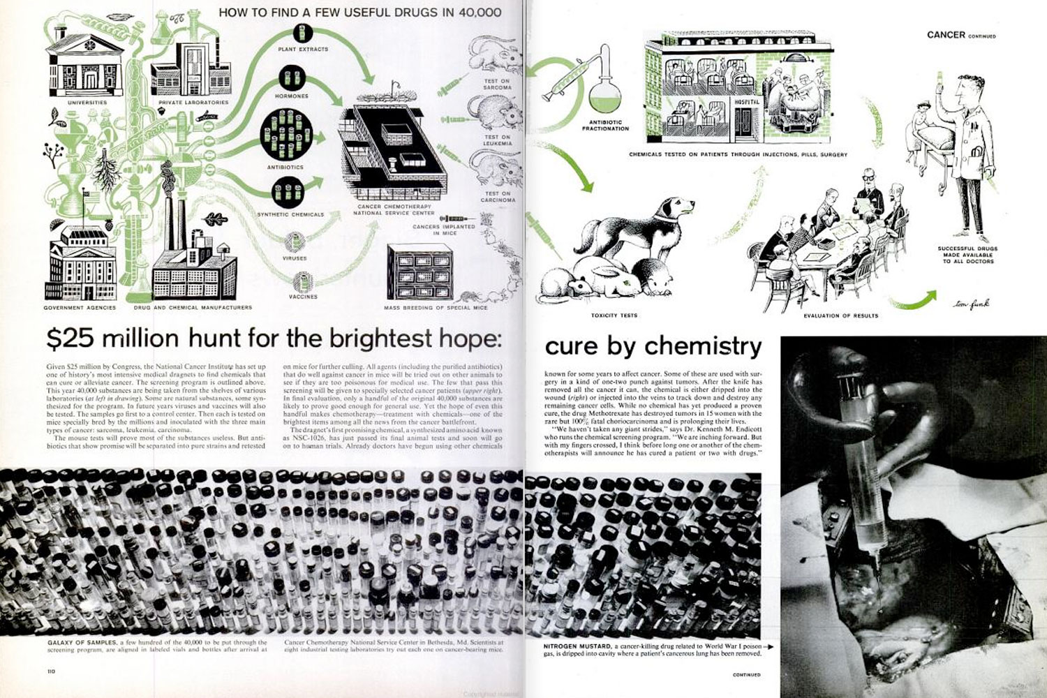
Caption from LIFE. "Learning how to tell, 1,000 women in Elmhurst, Illinois, watch American Cancer Society's film on detection routine, Breast Self-Examination. In seven years 6.5 million women have seen it." NOTE: This image is a composite of two photos, with the "onscreen" picture literally taped on to the other of the film audience.Al Fenn—Time & Life Pictures/Getty ImagesA movie theater showing the American Cancer Society's film on detection routine, Breast Self-Examination.Al Fenn—Time & Life Pictures/Getty ImagesWomen learn about cancer, 1958.Peter Stackpole—Time & Life Pictures/Getty Images"Ladies' line-up forms in Bry's department store, Memphis, Tenn., for cervical smear test. Of 114 Bry employees taking test, one had cancer. Memphis is the first U.S. city to start mass testing."Peter Stackpole—Time & Life Pictures/Getty ImagesCancer patient, 1958.Peter Stackpole—Time & Life Pictures/Getty ImagesStudying cancer patient, 1958.Peter Stackpole—Time & Life Pictures/Getty ImagesWomen study cancer cells under microscopes, 1958.Peter Stackpole—Time & Life Pictures/Getty ImagesLab conducting cancer research, 1958.Peter Stackpole—Time & Life Pictures/Getty ImagesCancer researcher, 1958.Al Fenn—Time & Life Pictures/Getty ImagesCaption from LIFE. "Nitrogen mustard, a cancer-killing drug related to World War I poison gas, is dripped into cavity where a patient's cancerous lung has been removed."Al Fenn—Time & Life Pictures/Getty ImagesCancer research, 1958.Al Fenn—Time & Life Pictures/Getty ImagesCaption from LIFE. "World's top expert on malignant cells, Dr. George Papanicolaou of New York's Cornell Medical College, displays projected cell images. He devised smear test that saves thousands of women."Walter Sanders—Time & Life Pictures/Getty ImagesCaption from LIFE. "At men's shelter, social worker Rubin Blum recruits men from Bowery for cancer diagnosis project. Of 800 men diagnosed, 90 had prostate cancer, 72 of these early, curable cases."Walter Sanders—Time & Life Pictures/Getty ImagesTesting for prostate cancer at the Francis Delafield Hospital in New York, 1958.Walter Sanders—Time & Life Pictures/Getty ImagesCancer researchers and patient at Francis Delafield Hospital, 1958.Walter Sanders—Time & Life Pictures/Getty ImagesCaption from LIFE. "Prostate x-rays of Bowery men ... are studied before operation at Francis Delafield Hospital to weed out incurable cases."Walter Sanders—Time & Life Pictures/Getty ImagesDoctors at Ohio State Penitentiary inject living cancerous cells into a prisoner's arm to test natural immunity, 1958.Peter Stackpole—Time & Life Pictures/Getty ImagesCaption from LIFE. "Injecting cancer cells into prisoners is done in immunity study." Ohio, 1958.Peter Stackpole—Time & Life Pictures/Getty ImagesDoctors at Ohio State Penitentiary inject living cancerous cells into a prisoner's arm to test natural immunity, 1958.Peter Stackpole—Time & Life Pictures/Getty ImagesCancer research on Ohio State Penitentiary prisoners, 1958.Peter Stackpole—Time & Life Pictures/Getty ImagesCancer research on Ohio State Penitentiary prisoners, 1958.Peter Stackpole—Time & Life Pictures/Getty ImagesDoctors at Ohio State Penitentiary remove growth of pre-injected cancerous cells from a prisoner's arm, 1958.Peter Stackpole—Time & Life Pictures/Getty ImagesCaption from LIFE. "Diseased cells, enlarged 2,000 times, are studied by Dr. George Z. Williams at National Cancer institute, Bethesda, Md., on ultraviolet TV microscope he helped develop. It reveals enzyme action."Andreas Feininger—Time & Life Pictures/Getty ImagesLIFE magazine, May 5, 1958. Best viewed in Full Screen mode. See button at right.LIFE MagazineLIFE magazine, May 5, 1958. Best viewed in Full Screen mode. See button at right.LIFE MagazineLIFE magazine, May 5, 1958. Best viewed in Full Screen mode. See button at right.LIFE MagazineLIFE magazine, May 5, 1958. Best viewed in Full Screen mode. See button at right.LIFE MagazineLIFE magazine, May 5, 1958. Best viewed in Full Screen mode. See button at right.LIFE MagazineLIFE magazine, May 5, 1958. Best viewed in Full Screen mode. See button at right.LIFE Magazine