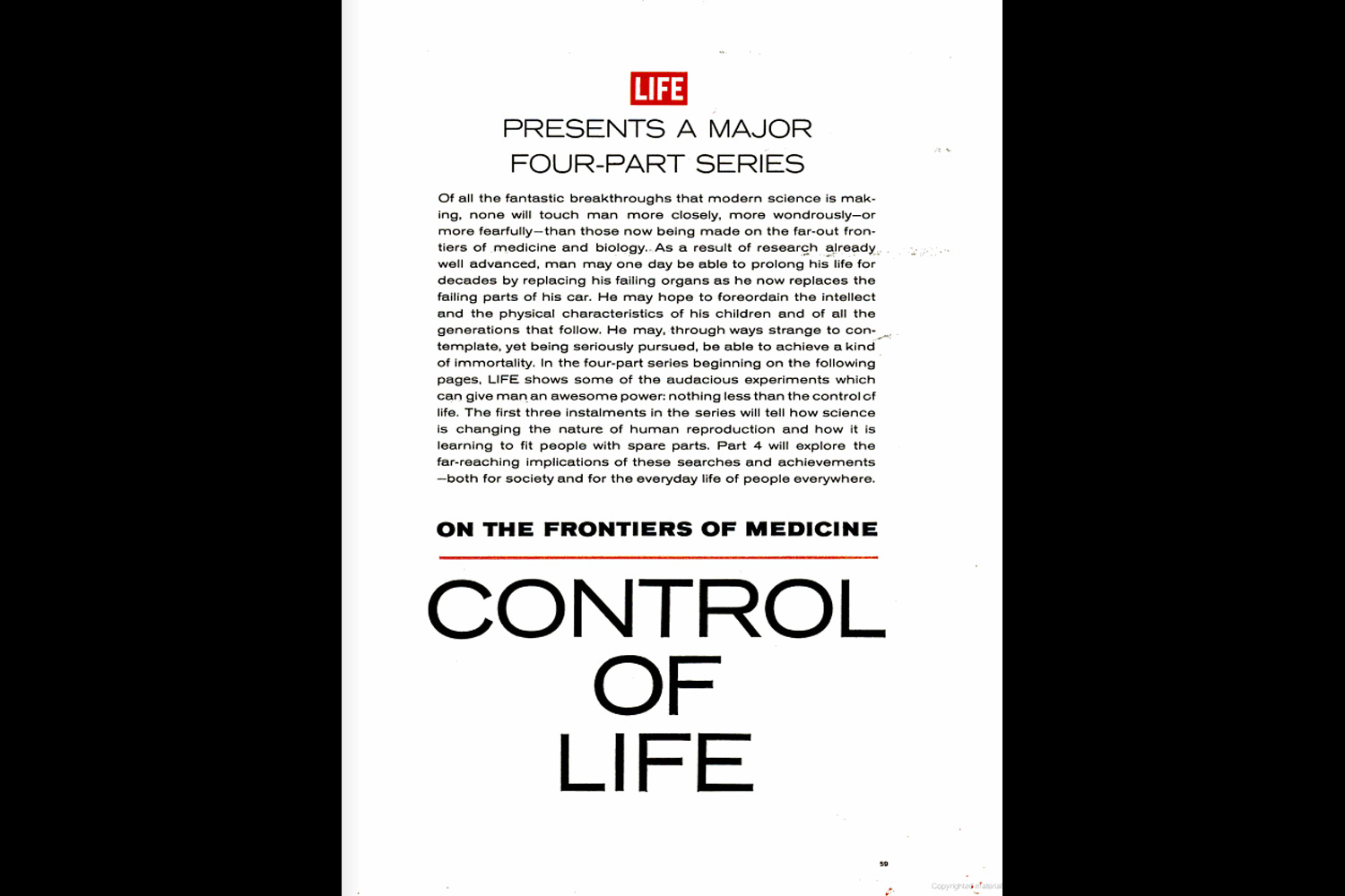
Control of Life: Pictures From a Medical Revolution, 1965
Control of Life: Pictures From a Medical Revolution, 1965
2 minute read
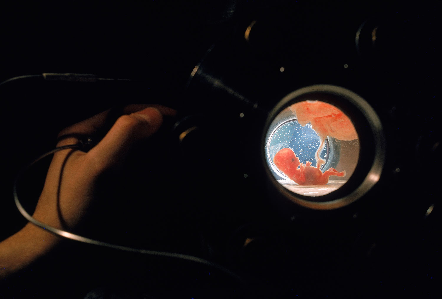
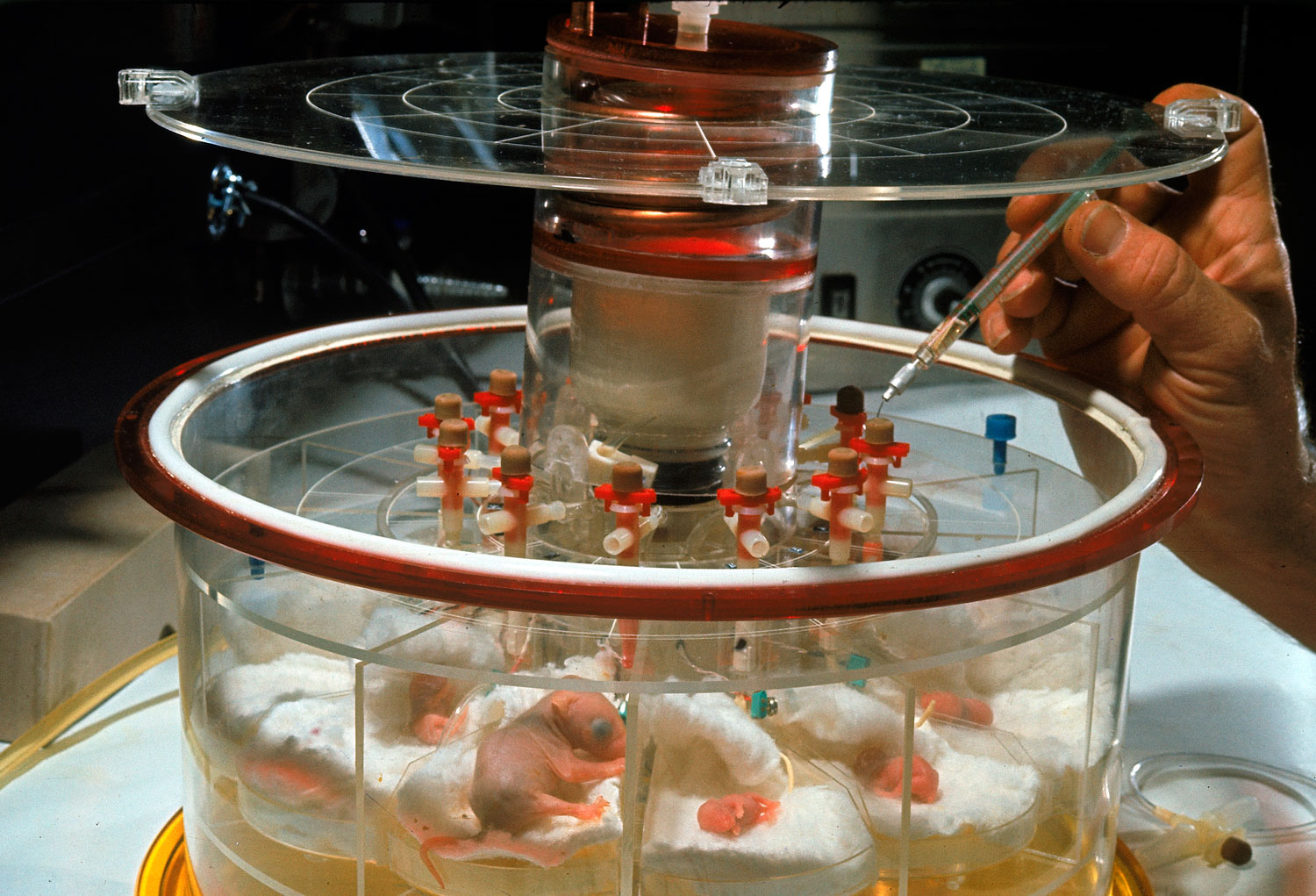
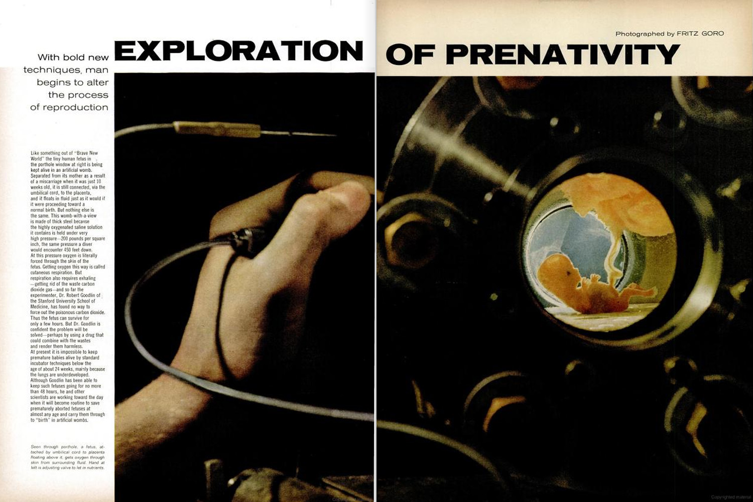
Caption from Life. "Seen through porthole, a fetus, attached by umbilical cord to placenta floating above it, gets oxygen through skin from surrounding fluid. Hand at left is adjusting valve to let in nutrients."Fritz Goro—Time & Life Pictures/Getty Images
In September 1965, LIFE magazine presented its millions of readers with a lengthy, thoughtful feature that it characterized as the first in an ambitious four-part series “on the profound and astonishing biological revolution.” Titled Control of Life, the series sought to create a framework within which LIFE could grapple with some of the most exciting and troubling recent (at the time) advances in science and medicine. The magazine described the intent of the four-part series in these words:
Of all the fantastic breakthroughs that modern science is making, none will touch man more closely, more wondrously — or more fearfully — than those now being made on the far-out frontiers of medicine and biology. As a result of research already well-advanced, man may one day be able to prolong his life for decades by replacing his failing organs as he now replaces the failing parts of his car. He may hope to foreordain the intellect and the physical characteristics of his children and of all the generations to follow. He may, though was strange to contemplate, yet being seriously pursued, be able to achieve a kind of immortality.
In the four-part series beginning on the following pages, LIFE shows some of the audacious experiments which can give man an awesome power: nothing less than the control of life.
Today, as previously unthinkable medical advances — from prenatal whole-gene sequencing to non-invasive cancer screening to at-home HIV tests — become more routine (if no less mind-blowing), and as ethical questions swirl around entire fields of research, LIFE.com presents photos from that first installment in the Control of Life series, as well as page spreads from the issue itself.
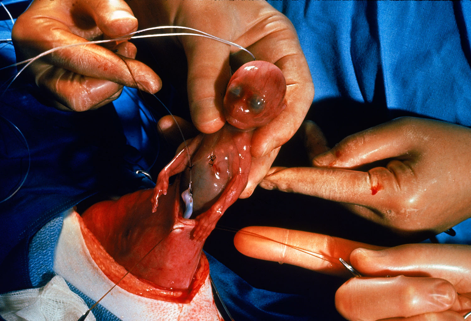
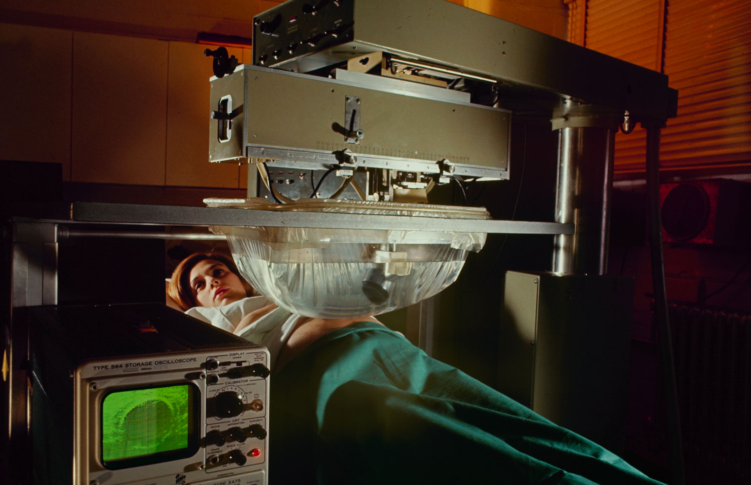
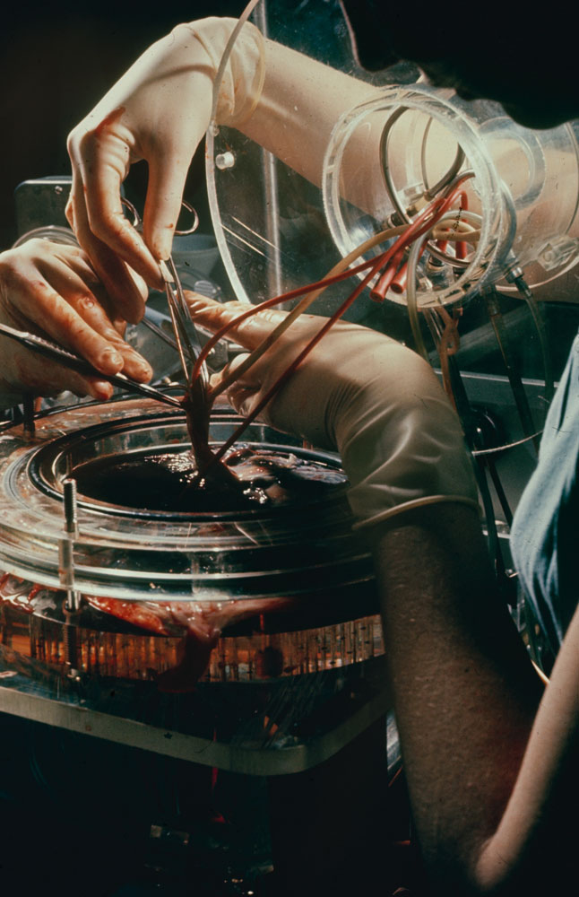
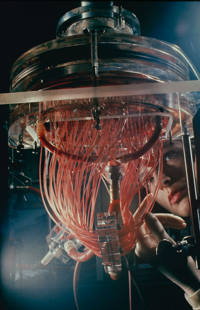
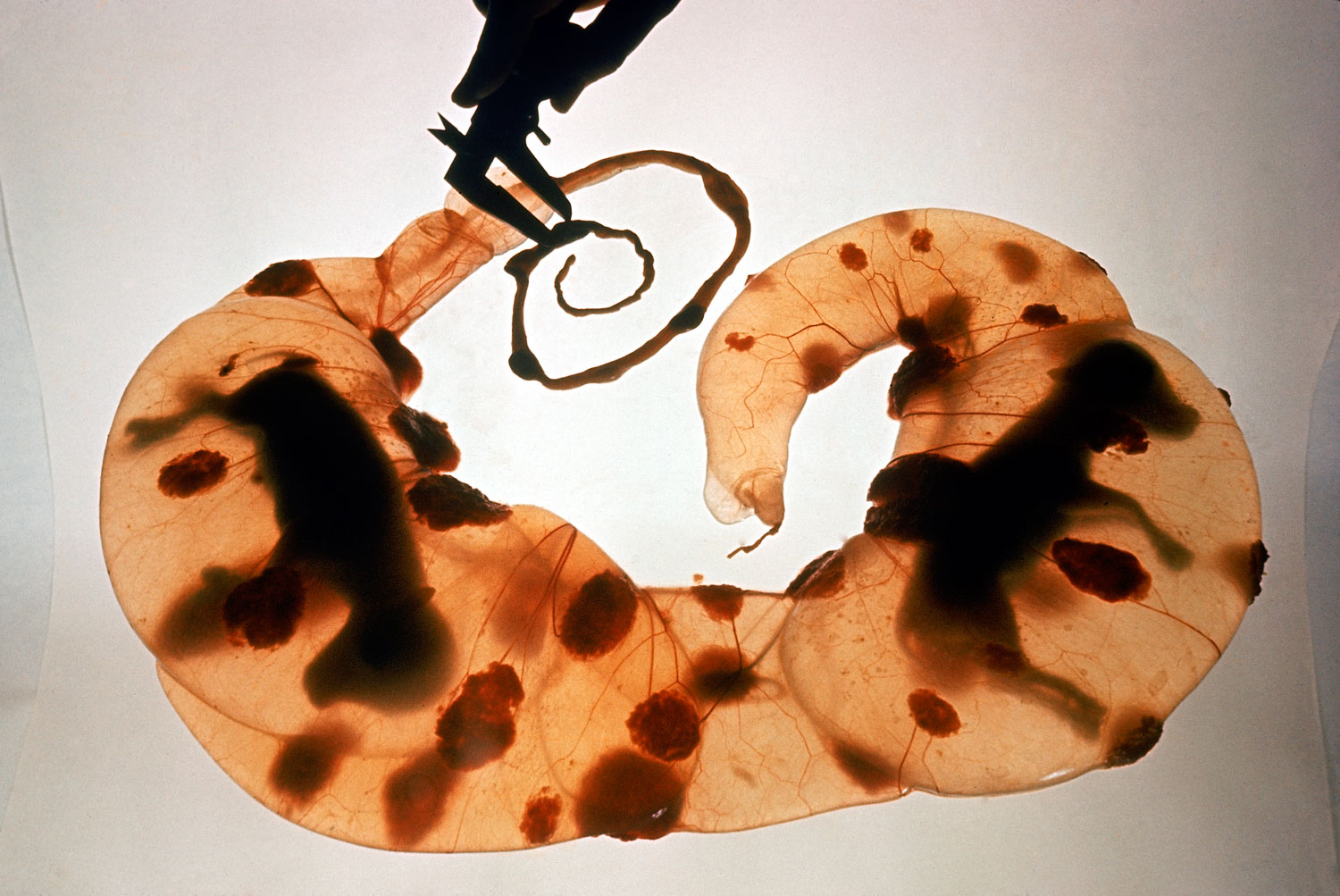
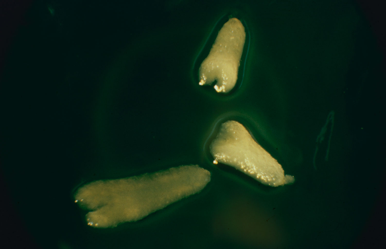
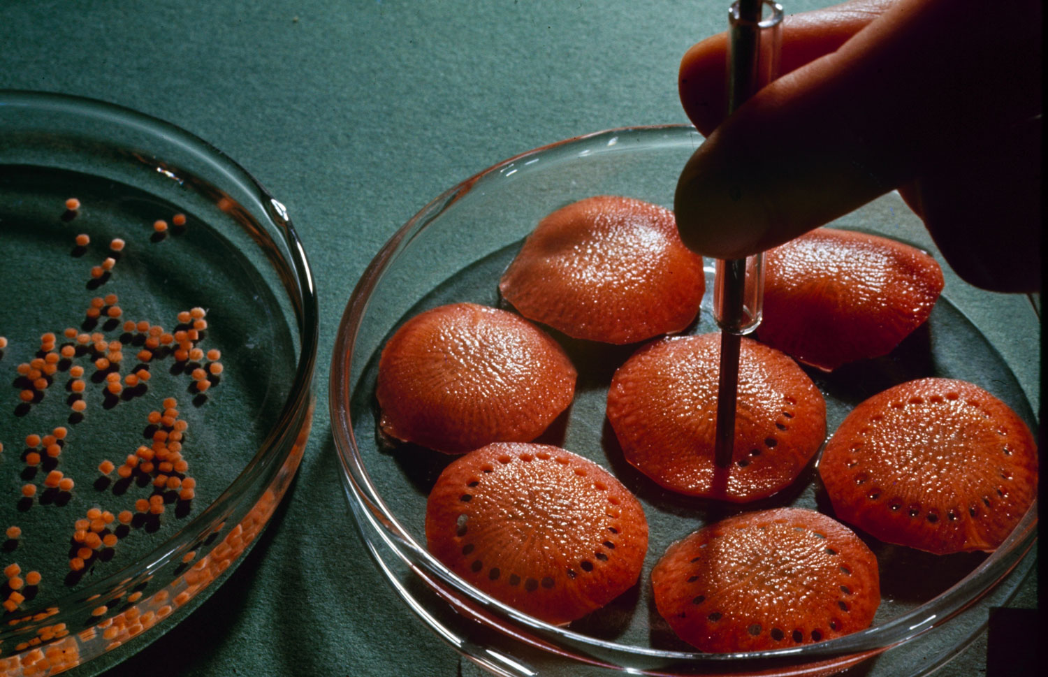
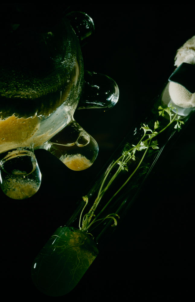
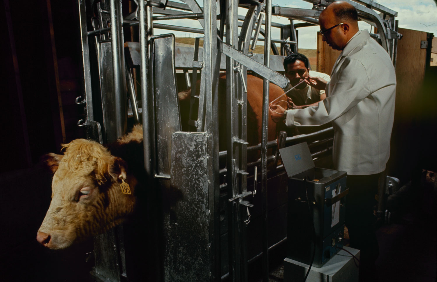
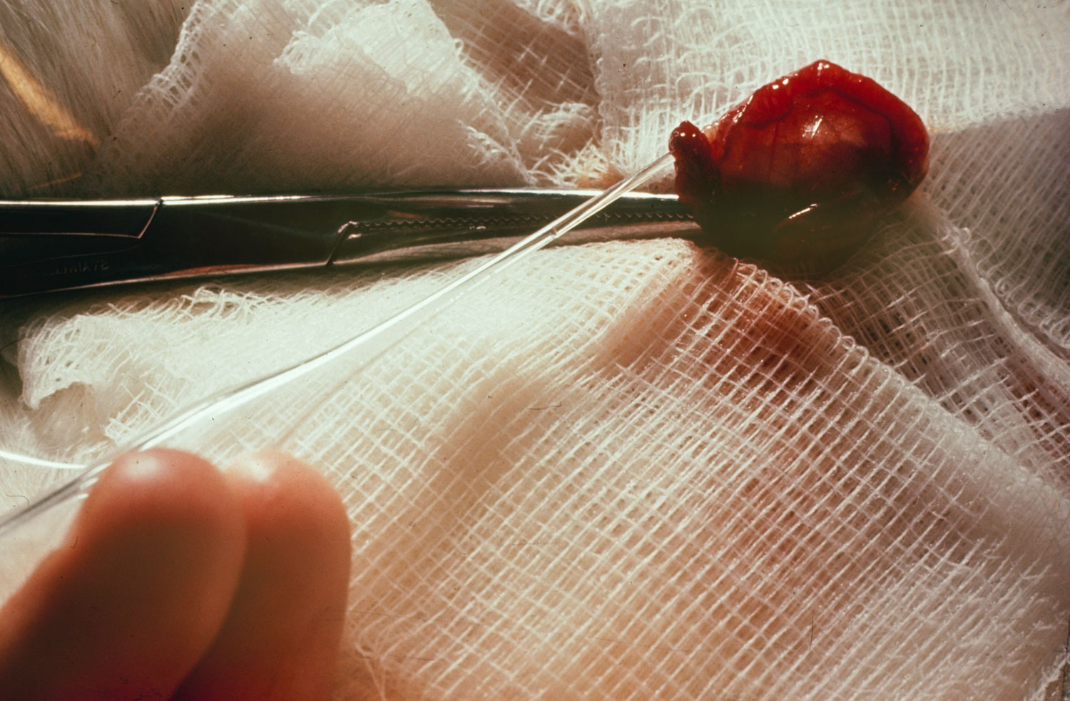
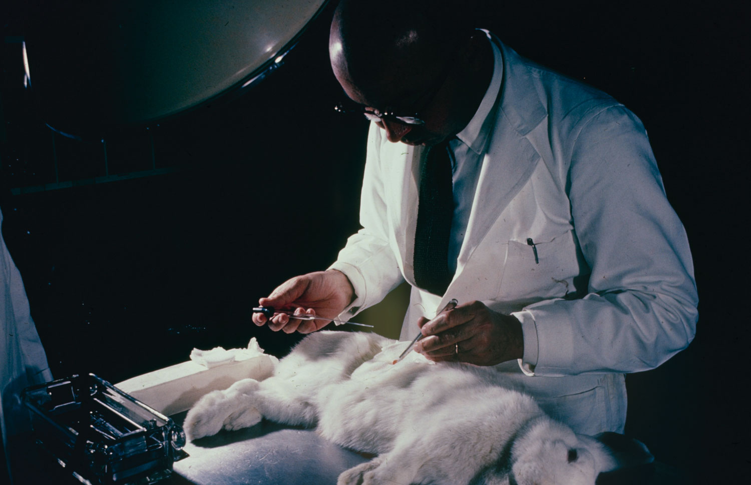
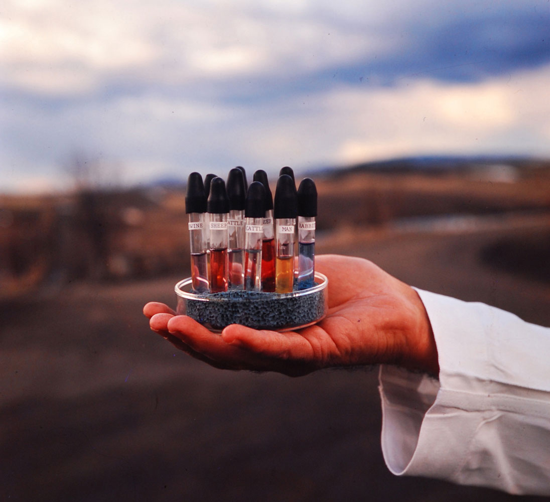
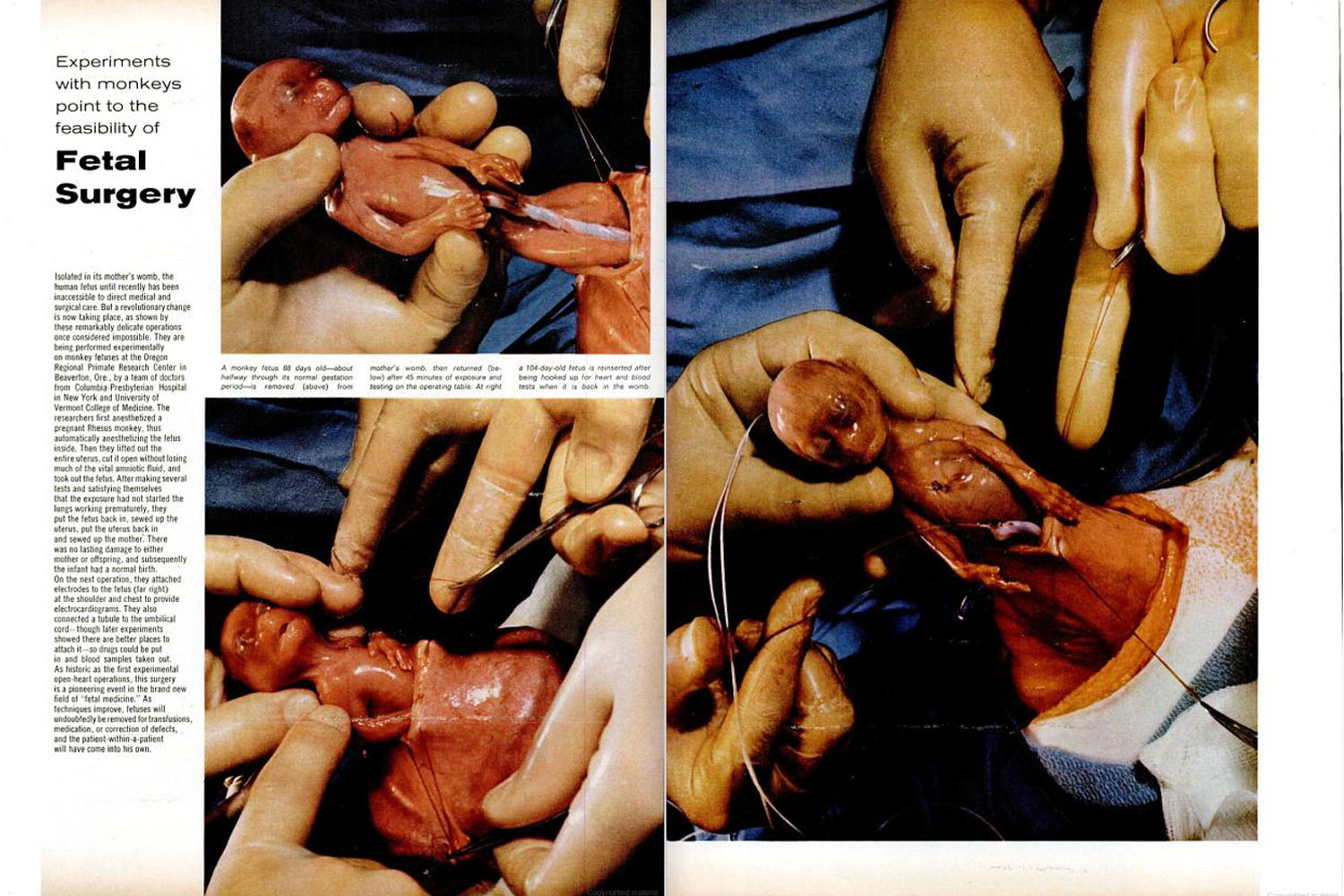
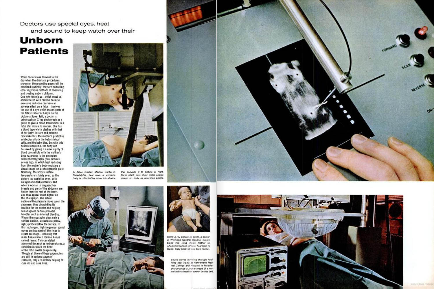
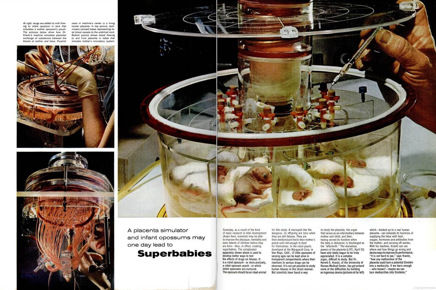
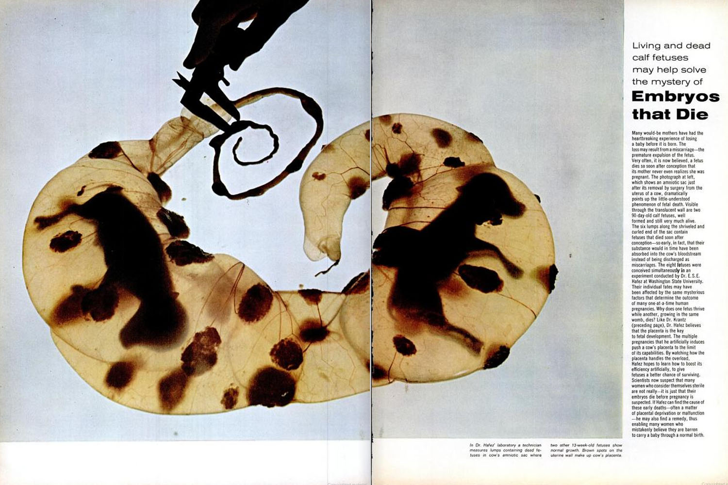
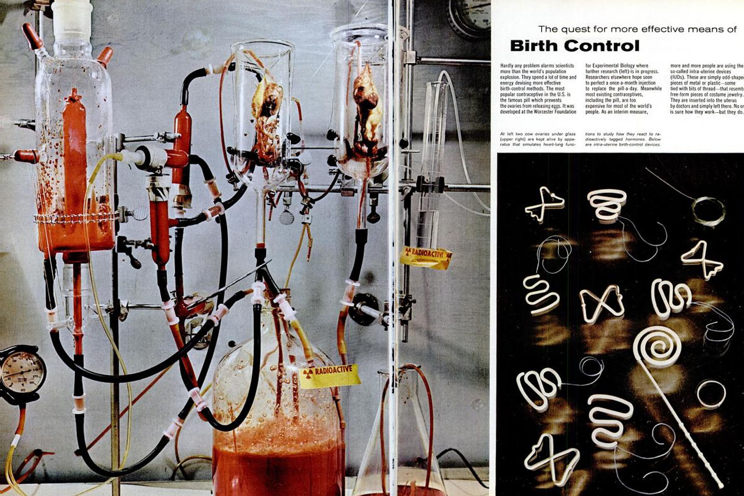
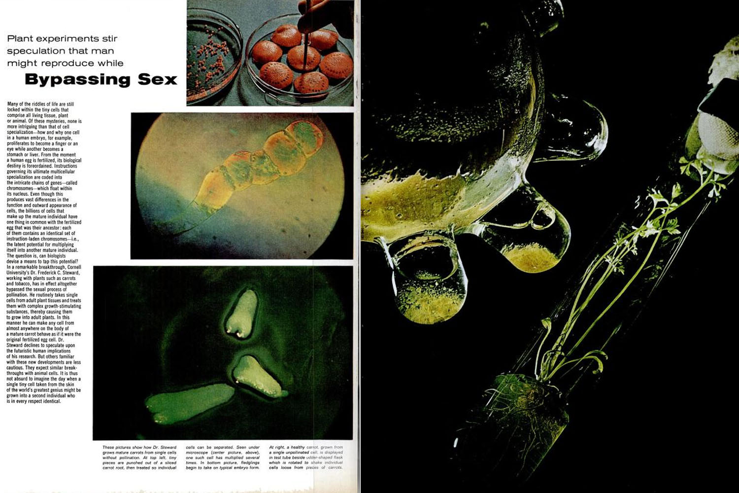
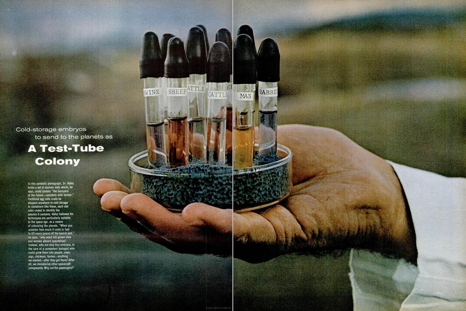
Caption from Life. "Seen through porthole, a fetus, attached by umbilical cord to placenta floating above it, gets oxygen through skin from surrounding fluid. Hand at left is adjusting valve to let in nutrients."Fritz Goro—Time & Life Pictures/Getty ImagesCaption from Life. "A 104-day-old [monkey] fetus is reinserted after being hooked up for heart and blood tests when it is back in the womb."Fritz Goro—Time & Life Pictures/Getty ImagesCaption from Life. "Sound waves traveling through fluid-filled bag at Hahnemann Medical College and Hospital in Philadelphia produce a profile of a normal baby's head on screen beside bed."Fritz Goro—Time & Life Pictures/Getty ImagesA machine simulates placental exchange of mother and fetus.Fritz Goro—Time & Life Pictures/Getty ImagesCaption from Life. "Drugs are added to milk flowing to infant opossum in tank that simulates a mother opposum's pouch."Fritz Goro—Time & Life Pictures/Getty ImagesBlood flows to and from placenta in tubes that simulate mother's circulatory system."Fritz Goro—Time & Life Pictures/Getty ImagesCaption from Life. "A technician measures lumps containing dead fetuses in cow's amniotic sac where two other 13-week-old fetuses show normal growth. Brown spots on the uterine wall make up cow's placenta."Fritz Goro—Time & Life Pictures/Getty ImagesCaption from Life. Intra-uterine birth-control devices. **please note, this image ran in color in the story - our archive only has a black and white version.Fritz Goro—Time & Life Pictures/Getty ImagesCarrots grow from single cells without pollination. Seen under microscope, fledglings begin to take in typical embryo form.Fritz Goro—Time & Life Pictures/Getty ImagesCarrots grow from single cells without pollination. Tiny pieces are punched out of a sliced carrot root, then treated so individual cells can be separated.Fritz Goro—Time & Life Pictures/Getty ImagesA healthy carrot, grown from a single unpollinated cell is displayed in test tube beside udder-shaped flask which is rotated to shake individual cells loose from pieces of carrots.Fritz Goro—Time & Life Pictures/Getty ImagesAn embryo calf from one cow is put into another.Fritz Goro—Time & Life Pictures/Getty ImagesCow embryos injected through a tube into the uterus of a rabbit. The uterus is attached to the rabbit which is covered by gauze. Fritz Goro—Time & Life Pictures/Getty ImagesCow embryo transferred into rabbit.Fritz Goro—Time & Life Pictures/Getty ImagesA set of dummy vials which "could contain "he barnyard of the future -- complete with farmer."Fritz Goro—Time & Life Pictures/Getty ImagesPage layout from September 15, 1965, issue of LIFE.Life MagazinePage layout from September 15, 1965, issue of LIFE.Life MagazinePage layout from September 15, 1965, issue of LIFE.Life MagazinePage layout from September 15, 1965, issue of LIFE.Life MagazinePage layout from September 15, 1965, issue of LIFE.Life MagazinePage layout from September 15, 1965, issue of LIFE.Life MagazinePage layout from September 15, 1965, issue of LIFE.Life MagazinePage layout from September 15, 1965, issue of LIFE.Life MagazinePage layout from September 15, 1965, issue of LIFE.Life MagazinePage layout from September 15, 1965, issue of LIFE.Life Magazine